A tour of the cell
How cells can be seen ?!
Light Microscopes ( LM )
Visible light passes through a specimen then and then through glass lenses, which magnify the image.
Were the first microscope.
Can magnify up to 1000 times.
Can't see the organelles with it.
Use to see: Most plant and animal cells, Nucleus, and Most bacteria.

LM ( Paramecium )
Magnification is the increase in the apparent size of an object.
* Paramecium is Protist.
Electron Microscopes ( EMs )
Used to study sub-cellular structures.
Instead of light, the EM uses a Beam of electron.
Can resolve biological structures as small as 2 nano-meters.
Can magnify up to 100,000 times.
Use to see: Viruses, Ribosomes, Atoms.
Two types of EM
Scanning Electron Microscope ( SEMs )
Focus a beam of electrons onto the surface of a specimen.
Providing image that look 3D.

SEMs ( Paramecium )
Transmission Electron Microscope ( TEMs )
Focus a beam of electrons through a specimen.
Used to study the cell internal structure.

TEMs ( Paramecium )
* Most cells are between 1 and 100 micrometers.
The quality of an image depends on
1- Magnification, the ratio of an object’s image size to its real size.
2- Resolution, the measure of the clarity of the image, or the minimum distance of two distinguishable points.
3- Contrast, visible differences in parts of the sample.
Cells
Prokaryotic, Eukaryotic,
All cells have several basic features in commen
1- Bounded by a plasma membrane.
2- Have one or more Chromosomes, carrying genes made of DNA.
3- Contain Ribosomes, tiny structures that make proteins according to instructions from the genes.
4- The interior of both types of cell is called the cytoplasm.
Prokaryotic

1- No nucleus.
2- DNA in an unbounded region called Nucleoid.
3- No membrane-bound organelles.
4- The ribosomes smaller and differ somewhat from Eukaryotes.
5- Cell wall: rigid structure outside the plasma membrane.
6- Flagella: locomotion organelles of some bacteria.
* Only organisms of Domain Bacteria and Archaea.
Eukaryotic

Animal Cell
Animal cells have ( Lysosomes & Centrioles ).
Lysosomes
Digestive Compartment
Lysosomes are digestive compartments within a cell.
A lysosome is a membranous sac of hydrolytic enzymes.
The enzymes and membranes of lysosomes are made by
Rough ER and processed in the Golgi apparatus.
A lysosome provides an acidic environment for its enzymes.
With the help of lysosomes a cell continually renews itself.
Many proteins engulf food particles into membranous sacs called food vacuoles.
Lysosomes fuse with food vacuoles and digest the food.
The nutrients are then released into the cell fluid.

It can digest marcomolecules such as
Proteins
Fats
Polysaccharides
Nucleic Acids
Also, use enzymes to recycle the cell's organelles.

The cells of people with inherited lysosomal storage diseases lack one or more lysosomal enzymes.
The lysosomes become engorged with undigested material,
eventually interfering with cellular function.
In Tay-Sachs disease, for example, a lipid digesting enzyme is missing, and the brain cells become impaired by an accumulation of lipids.

Plant Cell
Planet cells have ( Cell wall & Chloroplasts & Central Vacuole ).
* Planet cells have a rigid rather than thick cell wall.
( Fungi and many protists have rigid )
Cell walls protect cells and help maintain their shape.
Planet cells have Plasmodesmata are cytoplasmic channels
through cell walls that connect adjacent cells.
Vacuole
Maintenance of the cell
Vacuoles are large vesicles that have a variety of functions.
Paramecium has two contractile vacuoles.
Large central vacuole, which helps the cell grow in size by absorbing water and enlarging.
It also stockpiles vital chemicals and acts as a trash can.

Peroxisomes
Organelles in Plant and Animal cells
Peroxisomes are metabolic compartments that don't originate from the endomembrane system.
Its Function is
Detoxifying harmful compounds and making hydrogen peroxide (H2O2) By-product.
(H2O2) confined with in peroxisomes then converted to (H2O) by resident enzymes.
Break down fatty acids to be used as cellular fuel.

Eukaryotic Common Properties
1- DNA in a nucleus that is bounded by a membranous nuclear envelope.
2- Membrane-bound organelles.
3- Much larger than Prokaryotic.
4- Each organelles is bounded by a membrane with a lipid and protein composition that suits its function.

The structures and organelles of eukaryotic cells can be
organized into four basic functional groups as follows.
1- The nucleus and ribosomes carry out the genetic control the cell.
2- Organelles involved in breakdown or hydrolysis of molecules include lysosomes, vacuoles, and peroxisomes.
3- Mitochondria in all cells and chloroplasts in plant cells are involved in energy processing.
4- Structural support, movement, and communication among cells are the functions of components of the cytoskeleton, plasma membrane, and cell wall.
The endomembrane system
in Eukaryoric cells
Composed of Different membranes that are suspended in the cytoplasm within aneukaryotic cell.
These membranes divide the cell into Functional and Structural compartment (or Organelles).
Components of the Endo-Membrane system
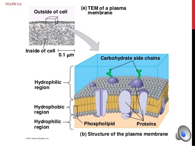
Plasma membrane
is selective barrier that
allows passage of Oxygen and Nutrients ( to cell ).
Drive out Wastes ( from cell ).
The general structure of a biological membrane is a double layer (bilayer) of
1- Phospholipids.
2- Protein.
3- Carbohydrates.

Plasma membrane structure
Nuclear Envelope
The nuclear envelope encloses the nucleus.
Nuclear envelope is a double membrane; each consist of a lipid bilayer ( double layer ).

lipid bilayer

Double Membrane
The nucleus envelope controls the flow of materials into and out of the nucleus.
The nucleus control cell activities.
By directing protein synthesis.
The nucleus contain most of DNA.
In the nucleus: DNA + Protein form Genetic Material called Chromatin.
Chromatin condenses to form Chromosomes.
The DNA is associated with many proteins in the structures called Chromosomes.

chromatin ti chromosome
The Protein helps organize and coil the long DNA molecule.
When cell is not dividing, this chromosome called chromatin.
The nucleolus is located within the nucleus.
and the site of ribosomal RNA (rRNA) synthesis.


Ribosomes ( Protein Factories )
Ribosomes are particles made of ribosomal RNA(rRNA) and protein.
Ribosomes are synthesized in the nucleolus, which is found in the nucleus.
Ribosomes carry out protein synthesis.
Cells that make a lot of proteins have a large number of ribosomes.
Some ribosomes are
Free (free ribosomes) suspended in cytoplasm
Bound (bound ribosomes) attached to the endoplasmic reticulum or the nuclear envelope.

Free and Bound Ribosomes
From DNA to Protein
Endoplasmic Reticulum
The endoplasmic reticulum (ER) accounts for more than half of the total membrane in many eukaryotic cells.
One of the major manufacturing sites in a cell is The Endoplasmic Reticulum.
There are two distinct regions of ER
1- Smooth ER: witch lacks ribosomes.
Functions
1- Synthesizes lipids.
2- Metabolizes carbohydrates.
3- Detoxifies poison.
4- Stores calcium ions.
* Our liver cells have large amounts of smooth ER.
2- Rough ER: with ribosomes attaching its surface.
Functions
1- Makes additional membrane.
2- Distribution of manufactured proteins.
3- A membrane factory for the cell.
The polypeptide is synthesized by a bound ribosome following the instruction of an mRNA.
Short chains of sugars are often linked to the polypeptide, making the molecule a glycoprotein.
Transport Vesicle, a vesicle that moves from one part of the cell to another.
The vesicle buds off the Rough ER membrane.
The vesicle now carries the protein to the Golgi Apparatus.
An important synthesis in the book page 43 ( Figure 3.6B ).

Smooth and Rough ER
Golgi Apparatus
Shipping and Receiving Center
Consist of flattened membranous sacs.
A cell may contain many, even hundreds of these stacks ( stack of flattened sacs ).
After leaving the ER, many transport vesicle travel to the Golgi Apparatus.
Note that: The flattened Golgi sacs are not connected. ( as are ER sacs ).
Functions of Golgi Apparatus
1- Modifies Products ( Protein + lipids ) of the ER.
2- Receive and Pack materials into transport vesicles.
Various Golgi enzymes modify the carbohydrates protein of the glycoprotiens made in the ER.
How? Removing some sugars and substituting others.
Watch (1:30 - 3:50) Golgi
Energy-Converting Oranelles
.svg/2000px-Animal_mitochondrion_diagram_en_(edit).svg.png)
Mitochondria
In all eukaryotic cells
Mitochondria are organelles that carry out cellular respiration.
nearly in all eukaryotic cells.
Converting the chemical energy of foods ( as sugar ) to the chemical energy.
The chemical energy of the molecule called ATP.
ATP = Adenosine Triphosphate.
ATP is the main energy source for cellular work.
It enclosed by two membranes, composed of
1- Phospholipids bilayer.
2- Unique collection of Protein.
The inner membrane is highly folded and contain many embedded proteins molecules that function in ATP synthesis.
The Folds called cristea, increase the membrane's surface area, enhancing the mitochondrion's ability to produce ATP.
The mitochondrion has two internal compartments :-
1- The intermembrane space.
The narrow region between the inner and the outer membranes.
2- The mitochondrial matrix. ( which contain )
1- Mitochondrial DNA and Ribosomes.
2- Many enzymes, that catalyze some of the reactions of cellular respiration.
The Common things between Mitochondria and Chloroplasts :-
They are not part of the endomembrane system.
They have a double membrane ( inner and outer ).
in between space called inner membrane space.
Both have a matrix.
Contain their own DNA.

Chloroplasts
In plant cells
Chloroplasts convert solar energy to chemical energy.
Chloroplasts are the photosynthesizing organelles of all photosynthetic eukaryotic.
Photosynthesis, the conversion of light energy from the sun to chemical energy of sugar molecules.
The compartments inside the inner membrane :-
1- Stroma, is a thick fluid which contain chloroplasts DNA and Ribosomes and many enzymes.
2- Thylakoids, is a network of interconnected sacs.
3- Granum, is each stack of thylakoids. ( plural = Grana ).
- Grana is the chloroplast's solar power packs.
- the sites where the green chlorophyll molecules embedded in thylakoid membranes trap solar energy.
Cilia and Flagella

Cilia and Flagella ( Paramecium )
Microtubeles cause cilia anf flagella to move.

Compare between Cilia and Flagella
Both cilia and flagella are made of microtubeles.
Extracellular Components

Cell Walls
In plant cells
Cell Wall distinguishes plant cells from animal cells.
The cell wall protect the plant cell, maintains its shape.
Plant cell walls are made of :-
1- Mainly of cellulose.
2- Polysaccharides.
3- Proteins.
Plant cell walls my have multiple layers :-
1- Primary cell wall : relativity then and flexible.
Which allows the growing cell to continue to enlarging.
2- Middle Lamella : then layer between primary walls of 2 adjacent cells.
3- Secondary cell wall : thick layer between plasma membrane and the primary cell wall.
In some cells.
Like ( Wood )
Plasmodesmata, the structure that connect two adjacent cells together.

Extracellular Matrix ( ECM )
Animal cells lack cell walls, but covered by Extracellular Matrix ( ECM ).
The ECM is made up of Collagen Fibers, which
(1)holds cells together.
(2)Protects the plasma membrane.
