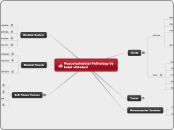
Musculoskeletal Pathology by hetal vithalani
Joints
connection between two bones solid joints are tightly connected to provide structural strengh Synovial joints have a joint space to allow for motion. 1. articular surface of adjoining bones is made of hyaline cartilage (type 2 collagen ) that is surrounded by a joint capsule synovium lining the joint capsule secretes fluid rich in hyaluronic acid to lubricate the joint and faciliate smooth motion.
Arthritis
Rhemotoid Disease
Chronic, Systemic autoimmune disease classically arise in women of late childbering age associatd with HLA - DR4- Hallmark is synovitis leag to formation of a pannus ( inflamaed granulation tissue) lead to destruction of cartilage and ankylosis joint clinical features arthrities with morning stiffness that improves with activitysymmertirc involement of pip joints of the finger Dip is usally spared. joint space narrowing, loss of cartilage, and osteopenia are seen on xray fever malaise weight loss and myalgias Rheumotoid nodules - central zone of necrosis surrounded by epithelioid histocytes arise in skin and visceral organ vasculitis - multiple organ may be involved baker cyst - swelling of bursa behind the knee pleural effusion lymphadenopathy and interestial lung fibrosis Mnumonic L – loss of joint spaceE – erosionsS – soft tissue swellingS – soft bones (osteopenia)Igm autoantibody against Fc portion of IGGneutrophils and high protein in synovial fluid complication invluede anemia of chronic disease and secondary amyloidosis
Osteoarthritis
noninflammatory progressive degeneration of articular cartilage primarily targeting weight-bearing joints (hip, knee, cervical/lumbar vertebrae), however the hands can also be affected — Heberden’s nodes at the DIP (distal interphalangeal) joint, Bouchard’s nodes at the PIP (proximal interphalangeal) joint.Joint stiffness and pain after inactivity—for example, morning stiffness after awakening, which generally lasts < 30 min.Joint stiffness and pain are aggravated by activity ∴ symptoms worsen as the day progresses, thus distinguishing osteoarthritis from rheumatoid arthritisCrepitus (feeling of crunching/crackling) when moving the jointOsteoarthritis in the fingers usually involves the DIP and PIP joints — vs. rheumatoid arthritis, which usually involves the MCP and PIP joints
infectious arthritis
Infectious Arthritis arthritis due to an infectious agent, uslly bacterial causes include N gonorrhoeae - young adults most common cause S. aureus older children and adults , 2nd most com cause Bacteria that are commonly found to cause septic arthritis are:Streptococci: the second most common causeHemophilus influenzae: was the most common cause in children but is now uncommon in areas where Hemophilus vaccination is practicedNeisseria gonorrhea: in young adults (although this is now thought rare in western Europe).Escherichia coli: in the elderly, IV drug users, and the seriously illcallsically involves a single joints, usally he knee presents are a warmjoint with limited range of motion fever increased white cell count and eleveted ESR are often present.
Seronegative Spondylorarthropathies
arthritis without ANA or rheumatoid factor (anti-IgG antibody), hence “seronegative”- strongly associated with HLA-B27- occur more commonly in malesSeronegative spondyloarthropathies include ankylosing spondylitis, reactive arthritis, and psoriatic arthritis.
Ankylosing spondylitis
chronic degenerative inflammatory arthritis primarily affecting the axial skeleton (spine, sacroiliac joints); also commonly affects peripheral joints (eg, shoulder, hip)Typical presentation:Young male in his 20s – 30s with chronic low back pain and stiffness that is:- worse with inactivity and in the morning- better with exercise, hot showers, and towards the end of the dayPatients also often have constitutional Sxs—eg, low-grade fever, fatigue, weight lossPathophysiology:1. Synovitis (inflammation of synovia) and enthesitis (inflammation of entheses—complex structures that attach ligaments and tendons to bone).2. At sites of synovitis/enthesitis there is bone destruction (mediated by osteoclasts, cathepsin K, metalloproteinases) and concomitant bone production with formation of syndesmophytes. Bone destruction → osteopenia or frank osteoporosis → ↑ risk of pathologic fracture—minimal trauma may cause:(a) lumbar spine fracture with resulting spinal cord injury (eg, cauda equina syndrome)(b) atlantoaxial subluxation due to odontoid fracture and/or transverse cervical ligament laxity3. Bony syndesmophytes eventually bridge gap between vertebrae → spinal fusion → rigid “bamboo” spine with decreased lumbar flexion (positive Schoberg test), obliteration of lumbar lordosis1. Anterior uveitis or iridocyclitis (confirm dx on slit lamp exam) → acute monocular pain, photophobia, blurry vision2. Aortitis, aortic root dilation → aortic regurgitation, which may progress to CHF (congestive heart failure)3. Cardiac conduction defects (eg, 3rd degree heart block)4. Involvement of thoracic spine → costovertebral rigidity and chest pain → ↓ chest expansion → restrictive pattern on pulmonary function tests
Reiter Syndrome
Can’t pee, Can’t see, Can’t climb a tree” to remember the classic triad of Reiter syndrome, a type of reactive arthritisongonococcal urethritis (Ureaplasma, Chlamydia):1. Urethritis — “Can’t pee”2. Inflammation of the eyes (conjunctivitis and/or uveitis) — “Can’t see”3. Inflammatory arthritis of large joints — “Can’t climb a tree”In addition to the classic triad above, patients with Reiter syndrome frequently develop:4. Oral ulcers5. Keratoderma blenorrhagica—erythematous scaly hyperkeratotic skin lesions on palms and soles6. Circinate balanitis—red scaly area on the glans penis with a gray, serpiginous annular edge that spreads outwards in phases
Psoriatic arthritis
asymmetric inflammatory polyarthritis that affects 10-30% of people suffering from psoriasis; usually involves small joints in the upper extremitiesWhen associated with psoriatic nail pathology (pitting of the nails), psoriatic arthritis commonly involves the DIP (distal interphalangeal) joints. Resorptive changes at the DIP joint with hyperemia (dactylitis) → involved fingers appear “sausage-shaped” and demonstrate a characteristic “pencil-in-cup” deformity on x-ray.The underlying process in psoriatic arthritis is inflammation ∴ 1st line therapy = anti-inflammatory agents like NSAIDs (eg, diclofenac, naproxen)
Gout
Buildup of uric acid (most commonly caused by underexcretion of uric acid) → crystals of monosodium urate are deposited in the articular cartilage of joints, tendons and surrounding tissues → gout
Acute gout
Acute Gout S/Sx:Excruciating, sudden, unexpected, burning pain, as well as swelling, redness, warmth, and stiffness in the affected joint.Mild fever; tachycardia, but only as a transient sympathetic response to the excruciating pain of an acute attackAcute gout most commonly involves the 1st MTP (metatarsophalangeal) joint—podagra = gouty arthritis of the 1st MTP.The extensor tenosynovium on the dorsum of the midfoot is also commonly affected during an acute gouty attack.
Chronic gout
If gout is poorly controlled, monosodium urate crystals may deposit in periarticular soft tissues → tophi: granulomas with multinucleated giant cells and negatively birefringent monosodium urate crystals (yellow when parallel to the slow ray)Brisk granulomatous reaction within tophi can destroy bone → erosive arthritisTophi may form around joints, but are also commonly found in earlobes, Achilles tendons, and olecranon and patellar bursae.
Primary Gout
Overproduction of uric acid may cause primary gout. For example:Alcoholism → ↑ turnover of adenine nucleotides (ie, ATP) during the conversion of acetate to acetyl-CoA → ↑ urate productionProtein-rich diets: red meat, seafood, beerUnknown enzyme defectsPrimary gout may also occur secondary to ↓ excretion of uric acid. For example:Thiazide diureticsLead poisoning → interstitial nephritis → ↓ urate excretionAlcoholism → ↑ production of lactate, which is an antiuricosuric agent because lactate competes with urate for the same renal excretion sites → ↓ urate excretion[Note: Alcohol can cause hyperuricemia by both increasing urate production and by decreasing urate excretion, as describe above]
Secondary Gout
Like primary gout, secondary gout can be caused by overproduction or underexcretion of uric acid.↑ nucleated cell turnover → overproduction of uric acid. Examples include:1. Cancer2. Psoriasis3. Treating leukemia; tumor lysis due to chemotherapyInborn errors of metabolism → overproduction of uric acid. For example, Lesch-Nyhan syndrome:1. X-linked recessive, complete HGPRT (hypoxanthine-guanine phosphoribosyltransferase) deficiency ∴ cannot re-use free purines via the salvage pathway2. HGPRT deficiency → ↑ PRPP (5-phosphoribosyl 1-pyrophosphate), which activates enzymes in the de novo purine synthesis pathway → ↑ de novo purine synthesis → hyperuricema, which may cause gout.3. The main clinical expression of Lesch-Nyhan syndrome is mental retardation and self-mutilation ∴ if gout occurs it is considered secondary gout.Chronic renal disease → ↓ excretion of uric acid
Psudo Gout
Usually affects the large joints, classically the knee. It usually affects people >50 years old and affects both sexes equallyUsing polarized light microscopy, calcium pyrophosphate dihydrate crystals:have rhomboid, square, or rodlike structuresare weakly and positively birefringent: think “blue when parallel, yellow when perpendicular”—i.e., the crystals are blue when the long axis of the crystal is parallel to the axis of slow rotation of light of the red plate compensator and yellow when the crystal is perpendicular to the axis of the compensator
Tumor
Nuromuscular Junction
Myasthenia Gravis
MG is caused by a Type II hypersensitivity reaction: autoantibodies bind postsynaptic acetylcholine receptors (AChR) → inhibition of ACh binding, internalization/degradation of AChRs, and complement-mediated postsynaptic membrane damage → ↓ number of functional AChRs and simplification of the postsynaptic membrane architecture → myasthenic muscle weakness, which:Worsens with muscle use (eg, exercise, high rate repetitive nerve stimulation) and is ∴ worse at the end of the day, improves with rest2. Fluctuates in the course of minutes/hours/days3. Usually begins with the extraocular muscles → ptosis and diplopia are the most common initial complaints
Lambert-Eaton Myasthenic Syndrome)
LEMS is believed to be caused by a Type II hypersensitivity reaction: autoantibodies bind presynaptic PQ-type voltage-gated calcium channels → ↓ fusion of synaptic vesicles with presynaptic membrane ∴ ↓ release of ACh into synaptic cleft ∴ less ACh is available to stimulate postsynaptic receptors → muscle weakness, which:1. Improves with muscle use2. Usually begins with proximal symmetric involvement of limbs, especially the proximal legs → difficulty climbing stairs or rising from a seated position, gait abnormalities
Skeletal System
Osteogenesis imperfecta
congentile defect of bone resorption resulting in structurally weak bone. most commonly due to an autosomal dominant defect in collegen type 1 synthesis multiple fractures of bone - please differenciate with child abuse - absent of brusing bule sclera thining of scleral colleen reveals underlying choroidal veins Hearining loss - bones of the middle ear easily fracture.
Osteopetrosis
Osteopetrosis inherited defect of bone resorpiton resulting in abnormally think, heavy bone that fractures easily Due to poor osteoclast function. multiple genetic variants exist carbonic anhydrase II MUTATION LEADS to loss of the acidic microenviroment required for bone resorption clnical features include bone fractures anemia thrombocytopenia luckopenia extramedullary hematopoiesis - due to replacement of bone merrow vison and hearing impairment - due to impingement on cranial nerves hydrocephalus - due to narrowing of the foramen magnum renal tubular acidosis - seen with carbonic anhydrase II mutation - lake of carbonic anhydrase results in decreased tubular reabsorption of HCO3- leading to metabolic acidosis treament is bone marrow transplant - osteoclasts are derived from monocytes.
Achondroplasia
impaired cartilage proliferation in the growth plate common cause of dwarfism Due to an activating mutation in fibroblast growth factor receptor 3 FGFRautosomal dominant Most mutations are sporadic and related to increased paternal age. clinical features short extremitles with normal sized head and chest - due to poor endochondral bone formation intramembranous bone formation is not affected. endochondral bone formation is charactrerized by formation of cartilage matrix, which is then replaced by bone it is the mechanism by which long bones growintramembramous bone formation ois characterized by formation of bone without a prextisting cartilage matrix it is the machanism by which flate bone develop
Vascular Necrosis
Avascular Necrosis ischemic necrosis of bone and bone mmarrow causes include trauma or fracture , steroids sickle cell anemia and caisson disease osteorthritis and fracture are major complications
Skeletal Muscle
Dermatomytosis
DM: antibody-mediated damage of blood vessels surrounding muscle fascicles (in the perimysial connective tissue) → perifascicular inflammation and atrophyDermatomyositis (DM) and Polymyositis (PM) are noninfectious inflammatory myopathies that present with symmetric proximal muscle weakness; normal reflexes, normal sensationBoth DM and PM are more common in women than men; peak incidence in adults 40 – 50 years of ageIn DM there is skin involvement; in PM the skin is not involved.Both DM and PM are associated with visceral malignancy, especially lung cancerSkin involvement in DMGottron’s sign: erythematous scaly eruption or dusky purple patches over the extensor surfaces of the knuckles (metacarpophalangeal and/or interphalangeal joints), elbows, and kneesHeliotrope rash (“racoon eyes”): lilac or violaceous eyelid discoloration with periorbital edemaShawl sign: erythema of the chest and shouldersV-sign: erythema in a V-shaped distribution over the anterior neck and chest
Polymyositis
cell-mediated (CD8 cytotoxic T cells and macrophages) damage of muscle fibers → endomysial inflammation (necrotic and regenerating muscle fibers scattered throughout the fascicle, not limited to the fascicle periphery as in DM)
X-linked muscular dystrophy
Degenerative disorder characterized by muscle wasting and replacement of skeletal muscle by adipose tissue Due to mutations of dystrophin dystrophin is important for anchoring the muscle cytoskeleton to the extracellular matrix mutations are often spontaneous large gene size predisposes to high rate of mutation.
Duchene Muscular dystrophy
Gene: Dystrophin, on X-chromosome → rarely affects girls1 in 3500 boysThe dystrophin links the actin cytoskeleton of smooth, cardiac and skeletal muscle cells to the extracellular matrix.Disease phenotype requires mutations at the reading frame level (deletion, nonsense, frameshift)Clinical course: pelvic muscle weakness that ascends
Becker Muscular Dystrophy
Becker’s muscular dystrophy: same gene is mutated, but symptoms are less severe
Soft Tissue Tumors
L
Lipoma
Lipoma = most common soft tissue tumor. Lipomas are benign tumors composed of fatty tissue → surgical excision is curativeClinical presentation:Lipomas present as masses which are:1. Soft to the touch2. Usually moveable3. Generally painless — angiolipomas are the only painful lipomasThere is a correlation between the HMG I-C gene (previously identified as a gene related to obesity) and lipoma development
Liposarcoma
Liposarcoma = most common soft tissue sarcoma. Liposarcomas are malignant tumors that arise in fat cells in deep soft tissue, such as that inside the thigh or in the retroperitoneum → will recur unless adequately excisedClinical presentation:- Middle-aged and older adults (usually ≥40 years of age)- Large bulky tumors which tend to have multiple smaller satellites extending beyond the main confines of the primary tumor.
Rhabdomyoma
Rhabdomyosarcoma
most common soft tissue tumor of childhood. RMSs are malignant tumors presumed to arise from primitive mesenchymal progenitors destined to form striated skeletal muscle cells.Clinical presentation:- Young children (usually <6 years of age)- Most commonly occur in the head and neck, but may also occur in the genitourinary tract — e.g., botryoid variant (sarcoma botryoides) = unique form of embryonal-type RMS that occurs in the bladder or vaginal wall in infantsAlthough most commonly the result of sporadic mutation, RMS may occur as part of certain inherited syndromes, including:1. Neurofibromatosis type 12. Li-Fraumeni syndrome3. Beckwith-Wiedemann syndromeDx: RMS is strongly suggested in a child that presents with a mass → biopsy demonstrating positive IHC (immunohistochemical) staining for muscle-specific proteins, including MyoD1, polyclonal desmin, muscle-specific actin, myosin, myogeninNote:1. MyoD1 = protein normally found in developing skeletal muscle cells that disappears after the muscle matures and becomes innervated ∴ MyoD1 serves as a useful IHC marker of RMS because MyoD1 is present in developmentally immature muscle (e.g., RMS) but is not found in normal, mature skeletal muscle.2. Myogenin = muscle-specific transcription factor essential for muscle development. Myogenin is expressed in developing skeletal muscle but not expressed in mature muscle ∴ myogenin is also a useful IHC marker of RMS.
