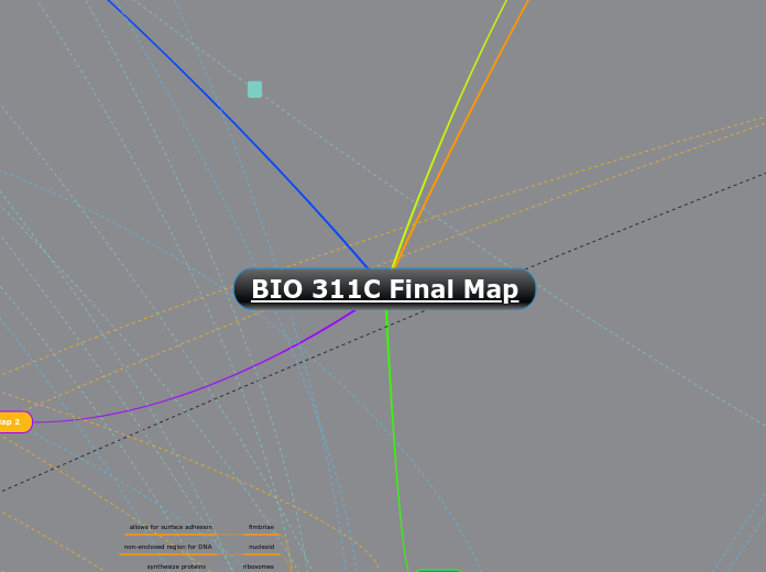
BIO 311C Final Map
Main topic
Map 4
Gene Regulation
Activators/Repressors are
Activated/Made
Protein
Binds to operator
Positive Regulation
Activator
Operon Gene Regulation On
Negative Regulation
Repressor
Operon Gene Regulation Off
Specific Transcription Factor
Last gene that enters the nucleus
Turns gene regulation on or off
Far Away from the Gene
Activator binds to enhancer
DNA bending brings activator
closer to promotion site
Activators bind to mediator proteins
Brings the activator closer to
the promotion site
Prokaryotes
Gene Organization
Structural genes
Genes whose expression is controlled together
Lac A
Structural Gene for B-Galactoside transacetylase
Lac Y
Structural Gene for B-Galactoside permease
Lac Z
Structural Gene for B-Galactosidase
Regulatory gene
Lac I
Codes for repressor protein
Regulatory regions
Promoter
Occurs structural and regulatory gene/s
Operator
“Switch” is a segment of DNA
Location where protein binds
Proteins are called activators/repressors
The binding causes positive/negative regulation
Turn on gene expression
Turn off gene expression
Regulation Through Operons
Occurs at level of transcription
Positive regulation= gene expression ON
expression at high level
With activator, transcription occurs
Without activator, no transcription occurs
Negative regulation= gene expression OFF
Repressor protein bond to operator sequence
With repressor, no transcription occurs
Without repressor, transcription occurs
Lac Operon
Example of negative and positive regulation
Regulation needs both repressor and operator
Lac= Lactose
Disaccharide made of glucose and galactose
Inducer of the lac operon
Lactose absent, repressor active, operon off
Transcription of structural genes is blocked
Negative regulation of operon
Lactose present, glucose scarce (cAMP level high)
Abundant lac mRNA synthesized
Operon ON: Induced/ high expression
Activator protein CAP is activated by cAMP
CAP helps RNAP to bind promoter
Facilitates transcription
Lactose present, repressor inactive, operon on
No glucose= operon ON
Inducible operon
All structural genes are transcribed
Forms a long mRNA
mRNA translates= B-Galactosidase, Permease, Transacetylase
Takes in more Lactose from outside
Break it down to glucose and galactose
Uses sugars as needed
Lactose present, glucose present (cAMP level low)
Little lac mRNA synthesized
Presence of Glucose operon OFF
Blocks Adenylyl Cyclase
Prevents production of cAMP
CAP cannot be activated
CAP can't help RNAP to bind promoter
Activators/Repressors involvement in regulation of gene expression
Eukaryotes
Transcription factors
Proteins that help turn specific genes "on" or "off"
Help increase or decrease level of transcription
General
Low levels of transcriptions
Background/basal
Bind to promoter and regions near
Specific
Change levels of transcription
Increase levels of transcription
Done by activators
High levels of transcription are reduced by repressors
Bind to distal control elements called enhancers
Present near or far from gene they are controlling
Control elements in DNA
Proximal
Sequences in DNA close to promoter
Bind general transcription factors
Distal
Enhancers
Sequences in DNA upstream or downstream of gene
Can be close or far from gene they are controlling
Bind to specific transcription factors
Prokaryotes
Operons
Helps with regulation with an on-off switch
"Switch"
Segment of DNA known as an "operator"
Positioned within promoter
Proteins bind to operators to turn on gene expression for multiple genes
OR to turn off expression
Postive regulation
Negative regulation
Eukaryotes
Gene Organization
Proximal
basal (general) expression
specific to eukaryotes
(ex. humans)
Distal
enhancers
bind activator proteins
activator bound to receptor is brought
to promoter via DNA bending proteins
transcription increased via
RNA polymerase II
Regulation through Operons
operons do not occur in eukaryotic cells
- made in mRNA w/ individual promoter
Transcription Factors
(instead of operons)
activators
repressors
Map 1
Map 3
Transcription and Translation
DNA
Structure
DNA strand
Sugar-phosphate backbone
Pentose (five-carbon) sugar (ribose)
Nitrogenous bases
Guanine
Adenine
Thymine
Cytosine
Chargaff's rule
A=T
G=C
Purines
A and G
Pyrimidines
T and C
Double stranded with complementary base pairing
Antiparallel
Monomer that makes DNA/RNA
Nucleotides
Components
Bonds present
Phosphodiester bond connects each nucleotide
Hydrogen bonds connect purines and pyrimidines
Pathways
Other Destinations
Organelles start in cytosol
Mitochondria
Convert energy for cellular respiration
Chloroplast
Produces various metabolites
Sensors of the external environment
Amino acids
Hormones
Vitamins
Lipids
Secondary metabolites
Nucleotides
Carries out photosynthesis
Peroxisome
Breakdown fatty acids through beta oxidation
Nucleus
Separates its contents from the cytoplasm
Regulate nuclear transportation
Holds the cell’s genetic material
Synthesizes the ribosome’s components
Cytoplasm
Translation takes place here
Endomembrane system
1-Free ribosomes enters Rough ER
2-Modified proteins in vesicles -> Golgi apparatus
3-Finalized vesicles transports to lysosome
4-Exocytosis occurs, protein exits cell
They undergo further modifications
Short chains of sugar molecules added/removed
Phosphate groups attached as tags
Proteins fold and undergo modifications
Addition of carbohydrate side chains
Secretory pathway (Polypeptide synthesis)
1-Begins on free ribosome
2-SRP binds to signal peptide
3-SPR binds to receptor protein
4-SPR leaves, synthesis continues
5-Signal peptide cleaved by enzyme
6-Finished polypeptide leaves ribosome
7-Folds into final conformation
Simultaneous translocation starts synthesis
This pauses snythesis
Examples of secreted proteins
Digestive enzymes
– Amylase
Peptide hormones
– Insulin
Milk proteins
– Casein
Extracellular matrix proteins
– Collagen
Serum proteins
– Albumin
Translation
Steps
match tRNA and amino acid
enzyme aminoacyl-tRNA synthetase
match tRNA anticodon with mRNA codon
mRNA
messenger RNA
nucleotides
genetic code
codons
three nucleotides code for amino acid
tRNA
transfer RNA
80 nucleotides long
anticodon
three amino acids
binds the tRNA to mRNA
amino acid attachment site
Ribosomes
rRNA
ribosomal RNA
proteins
facilitates coupling between tRNA and mRNA
Stages
Initation
mRNA and tRNA
1st amino acid attached to tRNA
Elongation
codon recognition
peptide bond formation
translocation
Termination
release factor
Transcription
DNA
RNA polymerase
binds to promoter
Initiation
polymerase unravels DNA
(downstream)
Elongation
RNA polymerase crosses
termination sequence
Termination
Prokaryotes
mRNA
DISTINCTIONS
no nucleus, transcription
takes place in cytoplasm
don't have introns; thus, when exiting,
doesn't have to keep mRNA stable
Eukaryotes
pre-mRNA
RNA processing:
- removes introns
- joins exons
mRNA
DISTINCTIONS
takes place in nucleus
CAP and TAIL
when exits nucleus, must
keep mRNA stable
uses RNA Polymerase II
(first needs to be binded by transcription factors)
Map 2
Cell Communications
Pathway of Signaling
Membrane receptors
G Protein Coupled Receptor
Signal molecule and GDP = Inactive
G-Protein alters shape, so GTP can bind to it
Now active, G-protein can activate enzyme (Adenylyl Cyclase)
Enzyme is activated at reception
Tyrosine Kinase Receptor
Polypeptides function as a Kinase
Kinase adds phosphates to other kinase through ATP
Tyrosine Kinase Receptor is activated
Processes cells use to make ATP
Cellular Respiration
Glycolysis
Location-
Cytoplasm/cytosol of the cell
Input-
2 ATP
1 Glucose
Output-
4 ATP
2 NADH
2 pyruvate
Net-
2 pyruvate
2 ATP
2 NADH
Substrate level oxidation-
ATP made
Pyruvate oxidation
Location-
Matrix of the mitochondria
Input-
2 Pyruvate
Output-
2 CoA
2 NADH
Net-
2 CoA
2 NADH
NO Substrate level oxidation-
ATP not made
Krebs Cycle / Citric Acid
Cycle
Location-
Matrix of the mitochondria
Input-
2 CoA
Output-
6 NADH
2 FADH2
2 ATP
Net-
6 NADH
2 FADH2
2 ATP
Substrate level oxidation-
ATP made
Oxidative phosphorylation
Location-
Inner mitochondrial membrane
Input-
O2
2 FADH2
10 NADH
Output-
H2O
28 ATP
Net-
H2O
28 ATP
Electron transport chain and chemiosmosis-
ATP made
Subtopic
Transduction Using cAMP
Transduction
Multi-step pathway that
helps amplify a signal
More coordination
More regulation
cAMP
Second messenger
Small, nonprotein, water-soluble molecules or ions
Used in signal transduction to
relay a signal within the cell
cyclic AMP
Formed from ATP
Synthesized using the enzyme Adenylyl Cyclase
Once used for the relay, cAMP is
converted into AMP by an enzyme
called phosphodiesterase
Cell Structures and Functions
Prokaryotic Cells
fimbriae
allows for surface adhesion
nucleoid
non-enclosed region for DNA
ribosomes
synthesize proteins
plasma membrane
structure; enclose
cell wall
rigid wall; provides structure
glycocalyx
capsule or slime layer outer coating
flagella
allows locomotion of organelles
bacterial chromosome
single, coiled chromosome
Eukaryotic Cells
Animal
centrosome
site for initiation of microtubules
lysosome
makes macromolecules
BOTH
flagellum
allows for movement
cytoplasm
gel-like substance; contains cell consistency
golgi apparatus
secretion of cell products & active in synthesis
peroxisome
metabolic functions
mitochondrion
regulates cellular respiration & produces majority of ATP
ribosomes
synthesize proteins
rough er
produce proteins
smooth er
synthesize proteins
plasma membrane
structure; enclose
cytoskeleton
support shape
nucleus
contains genetic material
Plant
central vacuole
storage and hydrolysis
cell wall
cell shape and protection
chloroplast
photosynthetic organelle
plasmodesmata
connects cytoplasms
R-group Orientation
Side Chain
Part of an amino acid
Electrically charged on basic and acidic groups
Four different groups
Polar
Tryptophan can function
Contains – OH, SH or NH groups
All contain polar covalent bonds
Hydrogen bonding
Non-Polar
Contains – H, CH or a carbon ring
Hydrophobic
All contain nonpolar covalent bonds
Van der waals forces
Basic
Hydrophilic
Positively charged
Acidic
Negatively charged
Ionic bonding
Disulfide bond
The only covalent bond
Interactions between R groups
Forms protein structures
Tertiary structure
Quaternary structure
Protein structures
Different levels
Primary structure
Covalent peptide bonds
Secondary structure
Hydrogen bonds
Tertiary structure
Electrostatic interactions/disulfide bridges
Modes of Transportation
Active Transport
Low to high concentration
Energy required
Passive Transport
High to low concentration
Diffusion
Facilitated Diffusion
Osmosis
No energy required
Membranes
Phospholipid Bi-layer
Hydrophilic heads and hydrophobic tails
Filter unwanted particles
Membrane fluidity
Saturated Fat
Less fluid membrane
Unsaturated Fat
More fluid membrane
Phospholipids in Membranes
Components
