Genitourinary System
RADIONUCLIDE CYSTOGRAPHY
voided (ml)x Residual counts/minutes over Max counts/min-Residual counts/minutes
-Patient to void before procedure
-Inject tracer mixed with saline into the tubing of the catheter
-Fill bladder to max fill limit
-Monitor P-scope carefully to view reflux, if reflux is visualized make note of amount of saline used at that time
-Image full bladder
-Deflate folley balloon and proceed to take post void images
-Record the amount of urine output
-Determine residual bladder volume
120 second immediate posterior void static image
Full Bladder Phase: 120 second immediate static of posterior and right/left posterior obliques
Filling Phase: Dynamic at 5 seconds for 1 minutes
Use Max Filling Formula to determine how much to fill the bladder: (age+2)x30= volume of bladder in mL
-Hand 500mL bag of normal saline at least 25cm above table
Patient Position: SUPINE with camera posterior
or sitting with back and pelvis against camera
99mTc-Sulfur Colloid
99mTc-DTPA
99mTc-Pertechnetate
Tracer is injected into the tubing connected to the catheter
Method of Administration: Catheter
Dose: 0.5-1.0mCi
-Cover imaging table with absorbent paper
-Patient to void before imaging
-Written consent for catherization
-Keep track of amount of saline from start to finish
Pregnancy
Evaluation and detection of vesicoureteral reflux (VUR)
MORPHOLOGICAL RENAL IMAGING
-Patient needs to void before imaging
-Patient to be imaged 2-4 hours after injection
Static Images: Posterior, RAO, LAO, RPO, LPO
-500K total counts per image
-Pinhole- 100K or 5 minutes
Patient Position: SUPINE with kidneys in field of view
-Single or dual head gamma camera
-Full field view for adults
-Zoom field of view for pediatrics
-Low energy all purpose (LEAP) collimator
-OPTIONAL: Pinhole Collimator
99mTc-GH (Gluceptate)
Secreted by glomerular filtration and tubular secretion
99mTc-DMSA (Dimercaptosuccinic Acid)
-Taken up in renal cortex
Dose: 5mCi (adults) 50uCi/kg (children)
-Well hydrated
-Void prior to imaging
-IV
Pregnancy
Patient movement
Detection of pyelonephritis
Differentiation of renal mass from normal variant
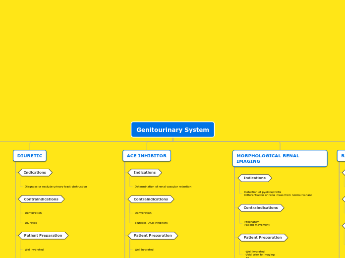
ACE INHIBITOR
A baseline renal scan needs to be performed before or after ACE scan to compare.
Final blood pressure taken (must be 70% of baseline blood pressure)
Blood Pressure taken every 15 minutes for 1 hour when given Captopril
25-50mg of Captopril given 1 hour before Tc99m or 40ug/kg of Enalaprilat given 10-15 minutes prior to Tc99m
ACE Inhibitors
ENALAPRILAT
10-15 minutes prior to radiopharmaceutical
40ug/kg in 10mL saline given over 3-5 minutes
CAPTOPRIL
Blood Pressure to be taken ever 15 minutes for 1 hour
1 hour prior to radiopharmaceutical injection
25-50mg pill given orally
Discontinue diuretics, ACE inhibitors, A2 receptors blockers- 4 DAYS PRIOR TO STUDY
NPO 4 to 6 hours prior to study
Patient to void before imaging
diuretics, ACE inhibitors
Determination of renal vascular retention
DIURETIC
Continue Dynamic acquisition for 20-30 minutes post furosemide injection
Diuretic: 40mg of Furosemide, injected slowly in IV at 20-30 minutes
Radiopharmaceutical and Interventional Pharmaceutical
Furosemide (LASIX)
Given 20-30 minutes post radiopharmaceutical
Pediatrics: 1mg/kg max dose of 40mg
Adult Dose: 40mg IV injection
99mTc- MAG3
-Excreted by tubular secretion -Rapid Plasma Clearance -Measures Effective Renal Plasma Flow (ERPF)
Dose: 10-15mCi
Serum creatine obtained
Well hydrated
Diagnose or exclude urinary tract obstruction
FUNCTIONAL RENAL IMAGING
Imaging
Processing
Renogram (time activity curve)
Region of interests (ROIs) are drawn around each kidney. Also, background regions are drawn (be careful to draw within body not outside the body), and also an aorta ROI is drawn around the aorta.
Procedure
Post Void: Posterior Static acquisition- 2 minutes
Function: Dynamic acquisition- 1 minute per frame for 30 minutes.
FLOW: Bolus Injection, 3 sec/frame for 1 min
Patient Position: SUPINE with kidneys and bladder in field of view.
Dose is used to measure that xiphoid process, pelvis/bladder and sides of body are with in field of view
Radiopharmaceutical
Equipment
-Single or dual head gamma camera
-Full field view for adults
-Zoom field of view for pediatrics
-Low energy all purpose (LEAP) collimator
99mTc-DTPA
-Glomerular filtration agent
-Measures Glomerular Filtration Rate
99mTc-MAG3
-Excreted by tubular secretion -Rapid Plasma Clearance -Measures Effective Renal Plasma Flow (ERPF)
Method of Administration: Intravenously
Dose: 10-20mCi
Patient Preparation
Start IV
Patient to void their bladder before imaging
Well hydrated-orally or intravenously
Contraindications
Diuretics
Dehydration
Assess renal function and urodynamics
Indications


