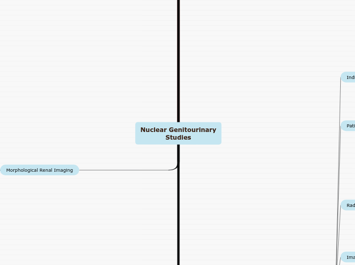
Nuclear Genitourinary
Studies
Vesicoureteral Reflux Study
abnormal
activity in upper urinary tracts during filling, at full capacity, and/or while voiding
all or nearly all solution voided from bladder
no reflux visualized
Total bladder volume residual post void volume and bladder volume at initiation of reflux can be measured
residual bladder volume [ml]= voided volume [ml] x residual counts/min
Divided by max counts/min - residual counts/min
Voiding phase
measure urine output
deflate foley and have patient void
place patient sitting on potty chair with camera posterior
Take 120 second immediate post void static and record CPM
Filling phase
once bladder is full, take 120 second immediate statics of posterior and L/R posterior obliques
monitor p-scope for signs of reflux (any activity above the bladder)
if reflux is seen, record amount of saline was infused at that time
fill until drip slows or voids around catheter
fill bladder to max capacity (age + 2) x 30 =volume instill [ml]
inject tracer into tubing connected to bladder catheter
image 5 sec/frame for 60 seconds
Posterior
patient positioning
sitting or supine
bladder and kidneys in FOV
MOL: compartmental with flow of urine
Critical organ: bladder (18-27 mrads/mCi)
indwelling bladder catheter
Dose: 0.5-1.0 mCi
Tc99m Sulfur Colloid
Tc99m pertechnetate
note amount of saline at start and finish
foley catheterization placement
inflate, balloon, tape to secure
aseptic technique
consent for catheterization
void prior to exam
Explain procedure to patient
Evaluation and detection of vesicoureteral reflux (VUR)
Diuretic Renal Imaging
Pre void and post void images of kidney and bladder
Lasix
Administer over 1-2 minutes by IV at 20 minutes
note frame number at time of administration
Dynamic
1-2 minutes/frame 20-30 minutes
Flow
30 frames x 2 seconds/frame
Generate Time Activity Curve
See Functional Renal Imaging
Patient Prep
SAME AS FUNCTIONAL RENAL IMAGING
Hydronephrosis
Renal obstructive neuropathy
Dehydration
Interventional Pharmaceutical
Furosemide
Pediatric dose: 1 mg/kg (Max 40 mg)
Dose: 20-80mg I.V.
Morphological Renal Imaging
Differentiate column of Bertin from mass
uptake in the column of Bertin, but not in a mass caused by tumor
Acute pyelonephritis
single or multiple defects resulting in decreased uptake
equal distribution in each kidney
smooth renal contour
Static images
Posterior/ RAO/LAO/RPO/LPO with kidney in center of FOV
2 hours post injection
Images
Data Acquisition
140 keV, 20% winodw
500k counts for each image
5 images total
LFOV gamma camera
SPECT: single, dual or triple head
Pinhole collimator for cortical images
prone preferred
parallel hole collimator for differential calculation
Positioning
include kidneys and bladder
Well Hydrated
void before imaging
Tc9m GH Gluceptate
Permits visualization of renal blood flow and imaging of the renal cortex
Secreted by Glomerular filtration and tubular secretion
Renal clearance is approximately 50% at 3 hours
Must be stored in the refrigerator
Child: 200 uCi/kg
Adult: 10-15 mCi
Intravenous
Tc99m DMSA
Child: 50uCi/kg
Webster's rule for pediatric dose: [age + 1] / [Age + 7] x adult dose
Adult: 5 mCi
10 mCI will give a higher dosimetry dose, but gives better image quality due to increased counts
25-50% of dose is in kidney in 1 hour and increases with time
Approx 16% will be in the urine in 3 hours after inj.
Taken up by renal cortex (proximal convoluted tubule) MOL tubular binding
90% binds to plasma proteins, preventing significant glomerular filtration
Tc99m dimercaptosuccinic
7 RADS to the renal cortex
Highest patient radiation dose of all renal imaging agents
pregnancy
not an absolute, risk-to-benefit ratio must be considered
patient movement
Confirmation of suspected hypertrophied column of Bertin
Scarring from acute pyelonephritis
pyelonephritis is usually results from reflux of infected urine
Adema
Renal Scintigraphy with ACE Inhibitor
Kidney Function decrease
Time to Peak activity decrease
prolonged renal parenchymal transit
Decreased renal uptake in one or both kidneys
Processing
ROI's
Background subtraction
cortical regions of the kidneys
Renogram Curve Analysis
Full FOV
Single or dual head camera
Posterior imaging
Obtain pre-void and post-void images
suspected ureteral obstruction
Serial Dynamic images
1-2 minutes/frame
20-30 minutes
Flow: Dynamic Acquisition
2-4 seconds/frame
60-120 seconds
Monitor patients blood pressure before administration of ACE inhibitor and every 15 minutes after for 1 hour
monitoring for hypotension
I.V. placement
Void before imaging
Fasting
4 hours fasting
Interventional Pharmaceuticals
Enalapirlat
Dose: 40ug/kg
I.V. over 3-5 minutes
Captopril
Given 1 hour before injection of radiopharmaceutical
Dose: 50mg
Pediatric dose: 0.5 mg/kg
Maximum: 25mg
P.O.
Crushed and dissolved in water
Radiopharmaceuticals
Tc99m
Energy: 140 keV
Tc99m DTPA
Target Organ: Kindeys
Tc99m MAG-3
Critical Organ: Bladder Wall
Target Organ: Kidneys
Pediatric: 0.15 mCi/kg
Maximum: 4mCi
Minimum: 1mCi
Adult: 5-10 mCi I.V.
No ACE inhibitors onboard
No diuretics onboard
Renal Hypertension
Renal Artery Stenosis
High Blood Pressure of unknown origin
Functional Renal
Imaging
Imaging
Results
Abnormal
asymmetric
retention of activity in the kidneys
Normal
reasonably symmetric
kidney activity peaking at 3-5 minutes and decreased to less than 50% by 20 minutes
Processing Images
Quantitative analysis - relative renal function
ROI's drawn around kidneys, abdominal aorta and background
Equipment
LEAP collimator
Full FOV for adults
Zoom FOV for pediatrics
Single or dual head gamma camera
Anterior imaging for renal transplants
Posterior imaging for native kidneys
Procedure
TOTAL TIME: 22 MINUTES
Post Void: Posterior static acquisition
2 minutes
Function: Dynamic acquisition
19 minutes of 20 s/frame; compressed to 1 min/frame
Flow: Dynamic acquisition
30 frames x 2 s/frame
Patient Positioning
Use dose to mark sternal notch in top 1/3 of image and bladder in bottom 1/3 of image. Use dose to ensure patients left and right sides are in the image
Supine
Patient Preparation
IV placement
Well hydrated
Bladder emptied immediately before imaging
Radiopharmaceutical
Energy
140 keV
Tc99m - MAG 3
Pediatric: 100 uCi/kg
Minimum 1 mCi
Functional, effective renal plasma flow agent
MOL: Tubular reabsorption/Glomerular filtration
Tc99m - DTPA
Target organ: Kidneys
Critical organ: Bladder
Dose
Pediatric: 200 uCi/kg
Minimum 2 mCi
Adult: 10-20 mCi
Functional glomerular agent
MOL: Glomerular filtration
Administration
Large antecubital vein preferred
Intravenous, bolus
Contraindications
Recent nuclear medicine studies
Pregnancy or breastfeeding
Indications
Postsurgical renal function assessment after pyeloplasty
Address renal function in infants with hydronephrosis
Assess for RAS or urinary reflux and scarring
Assess cause for acute renal failure or UPJ obstruction
Evaluate blood flow and function of renal transplant
Assess split renal function for native kidneys
