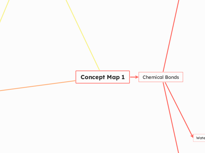
Concept Map 1
Chemical Bonds
Intramolecular Interactions
Dipole-Dipole
Ion-Dipole
Hydrogen Bonding
Van der Waals
Hydrophobic Interactions
Water Properties
Temperature Based
High Heat of vaporization
Expands when Freezing
Denser as a liquid
High Specific Heat
Structure Based
Universal Solvent
High Surface Tension
Hydrophobic/Hydrophilic Interactions
Intermolecular Forces
Covalent (electrons shared)
Electronegativity
>2.5
Polar
<2.5
nonpolar
Ionic (electrons transferred)
Eukaryotic/ Prokaryotic Cells
Domains of life
Archaea
Extremophiles
Extreme thermophiles
methanogens
Extreme Halophiles
Bacteria
Components
Nucleoid
Ribosomes
Plasma Membranes
Flagellum
Cell Wall
Fimbrae
Eukarya
Double Membrane Bounded
Animal Cells
Centrosome
region where microtubules are initiated
Flagellum
Lysosomes
Where macromolecules are hydrolized
Plant Cells
Chloroplast
converts energy of sunlight to chemical energy stored in sugar cells
Plasmodesmata
channels through cell walls that connects the cytoplasm of adjacent cells
Central Vacuole
used for storage, breaking down waste & hydrolysis of macromolecules
Cell Wall
maintains cell shape and protects cells from mechanical damage
Cells in plants and animals
Mitochondria
where cellular respiration occurs and ATP is generated
Plasma Membrane
Golgi Apparatus
responsible for the synthesis and secretion of a cells products
Cytoskeleton
microfilaments
microtubules
Endoplasmic Reticulum
Smooth ER
Rough ER
Nucleus
Nuclear Envelope
Nucleolus
Chromatin
Biomolecules
DNA/RNA
Polymer
Phosphodiester Linkage (5,3)
DNA= 2x strand
RNA= 1 strand
Monomer
Nucleic Acids
Pentane Sugar
Deoxyribose=DNA
Ribose=RNA
Nitrogenous Base
DNA
A,T,C,G
RNA
U
Phosphate Group
Proteins
Monomer
Amino Acids
Peptide Bonds
Structure
Primary
Secondary
Alpha + Beta Structures
H-Bonds
Tertiary
R group interactions
Disulfide Bonds
Quatenary
multiple tertiary protiens
Carbohydrates
Monosaccharides
Ketoses
Trioses (3C)
Pentoses (5C)
Hexoses (6C)
Aldoses
Disaccharides
Sucrose (glucose+fructose)
Lactose (glucose+galactose)
Maltose(glucose+glucose)
Polysaccharides
Storage
Starch (plants)
Amylose
Unbranched
a (1,4) glycosidic linkages
helical structure
Amylopectin
Branched
a (1,4), a (1,6) glycosidic linkages
helical structure
Glycogen (animals)
Extensively Branched
a (1,4), a (1,6) glycosidic linkages
helical structure
Structural
Cellulose
No branching
b (1,4) glycosidic linkages
linear structure
Lipids
Triglycerides (Fats)
Structure
Glycerol
fatty acids (3)
Saturated
No double bonds
solid at room temp.
Unsaturated
double bonds
liquid at room temp.
Phospholipids
Structure
glycogen
2 fatty acids
phosphate group
Function
forms phospholipid bilayers in cell membrane
amphipathic
Steroids
Structure
4 fused carbon rings
Cholesterol
High-Density Lipoprotein
helps remove excess cholesterol by taking it to the liver for excretion
Low-Density Lipoprotein
deposits extra cholesterol in blood cells which can lead to plaque buildup.
Hormones
Examples
Energy Investment Phase
trans & cis
Payoff Phase
Products: 2 pyruvate, 4 ATP, 2 NADH
Net Total: 2 pyruvate, 2 ATP, 2 NADH
x2
Inputs: 2 Acetyl CoA
Products: 2 ATP, 6 NADH, 2 FADH
Step 6 + 7: The two G3P become 2 pyruvate and 4 ATP, 2 NADH are produced.
Concept Map 2
Membranes
Tonicity
Osmosis (H2O moves to areas with higher concentration)
ability to cause cell to gain/lose water
hypotonic sol.
relatively lower concentration
compared to the cell
Plants: turgid (ideal)
animal: lysed
Isotonic sol.
same concentration as the cells
Plant: Flaccid
animal: normal (ideal)
Hypertonic sol.
relatively higher concentration compared to the cell
Plant: Plasmolyzed
animal: shriveled
Transport
Passive
diffusion down a concentration gradient w/out using energy
Facilitated
uses proteins and other channels to aid passive diffusion
Channels
channel that allows water/solute to enter (no shape change)
Ion Channels
ungated
constantly openn
gated
stretch
opens/closes when deformed
ligand
opens/closes when ligand binds to a receptor
voltage
opens/ closes when membrane potential changes
Carrier proteins
changes shape to move solute
Bulk Transport
Use vesicles to transport large molecules. membrane stretches to engulf particles
endocytosis
phagocytosis
takes in "food" particles
pinocytosis
takes in fluids
receptor mediated
uses receptors and ligands to take in molecules
cell takes in substances
exocytosis
cell ejects substances
Active
Uses energy to transport solute against its concentration gradient
electrogenic pump
creates a charge gradience generating voltage across a membrane
Proton Pump
Na/K Pump
1. 3 Na in the cytoplasm bind to the pump
2. phosphorylation via kinase triggered (PO4 attaches to the pump)
3. Pump changes shape and releases Na+ out
4. 2 K+ outside the cell attach to the pump while removing PO4
5. pump returns to its original shape
6. K+ comes off the pump and the cycle repeats
2 K+ in
3 Na+ out
Cells are slightly - because there are fewer positive charges in the cell
Phases:
Resting State: na and k voltage gated pumps are closed
Depolartization: Na pump opens allowing Na to flow in making it less negative (if this hits a threshold it moves on to the rising phase)
Rising Phase: cell becomes positive while K pumps are still closed until action potential is reached
Falling Phase: Some k pumps open while na pumps close making the cell overall negative
Undershoot: the cell uses na/k pump to help return it to the resting phase.
Cotransport
when active transport indirectly transport of another
sucrose/H+: H+ drives sucrose in when H+ is pumped out of the cell and a gradient is created
Permeability (high to low)
small, nonpolar > small, uncharged polar > large, uncharged polar > ions
Structure
Fluidity
Temperature
high temp= fluid
lower temp= rigid
saturated fats
lower diffusion+ rigid
Unsaturated fats
higher diffusion+ fluid
Cholesterol
helps regulated fluidity when too rigid or too fluid
phospholipid bilayer
Cellular Respiration
Glycolysis
with O2
pyruvate oxidation
Citric Acid Cycle
Oxidative Phosphorelation
Inputs: 10 NADH, 2 FADH
Chemosmosis
ATP Synthase
Here, H+ in the intermembrane space, go back down their concentration gradient. This energy is used to add an inorganic phosphate to ADP to form ATP
Electron Transport Chain:
Complex III
Complex IV
O2 + H+ = H20
Outputs: H2O, about 26 - 28 ATP
Complex II
FADH2 transfers electrons to complex II
Complex I
NADH transfers electrons to complex I
Q
Cyt c
Inputs: Acetyl CoA
Step 1: Oxaloacitate goes to an enzyme and with Acetyl CoA becomes Citrate.
Step 2: Citrate becomes Isocitrate.
Step 3: Isocitrate becomes alpha ketogluta and NADH is released.
Other Steps: 2 NADH, ATP, FADH are formed.
Products: 1 ATP, 3 NADH, 1 FADH
Inputs: 2 pyruvate
Products: 2 Acetyl CoA, 2 Co2, 2 NADH
Inputs: 1 glucose, 2 ATP
Step 1: glucose binds to hexokinase and with ATP becomes G6P.
Step 2: G6P becomes F6P
Step 3: F6P binds to the phospho-fructo-kinase enzyme and with ATP becomes fructose 1,6 bi-phosphate
Step 4 + 5: That fructose 1,6 biphosphate becomes 2 G3P
Without O2
Alchohol Fermentation
Inputs: 2 Pyruvate,
NADH
Outputs: Ethanol
NAD+
Lactic Acid Fermentation
Inputs: 2 Pyruvate,
NADH
Outputs: Lactate,
NAD+
Enzymes
Inhibition
Competitive Inhibition
An inhibitor binds to the active site of an enzyme. This doesn't allow for the substrate to bind and it stops the enzyme for functioning.
Non-competitive Inhibition
An inhibitor binds to a site other than the allosteric site, and changes teh enzyme's shape. Even if the substrate binds the active site, the ezyme no longer works.
Allosteric Regulation
Allosteric inhibitor
A regulatory molecule binds to the allosteric site of an enzyme an locks it in its in-active form. Substrates can't bind.
Allosteric activator
A regulatory molecule binds to the allosteric site of an enzyme an locks it in its active form. Substrates can bind.
Cooperativity
The binding of one substrate molecule to the active site of one subunit locks all other subunits into the active shape.
Feedback Inhibition
The end product of a metabolic pathway shuts down the pathway by going back to the first enyme and binding to its allopsteric site.
Cell Signaling
G Protein signaling pathway
Signal binds to the GPCR which changes its shape and activates it
GCPR binds to G protein, bound by GTP, which activates the G protein
Activated G protein binds to the adenylyl cyclase. GTP hydrolyzes which activates adenylyl cyclase and changes its shape.
Adenylyl cyclase converts ATP to cAMP
cAMP activates pka which leads to cell response
Aldosterone Pathway
Signal moves through the membrane
Signal binds to the receptor, changes its shape and activates the receptor
Active Receptor travels into the nucleus and binds to DNA
Transcription occurs which produces mRNA
mRNA leaves the nucleus, ribosomes bind and translation occurs, producing a protein.
Metabolism
Anabolic
pathway that consumes energy to build larger complex molecules
Endergonic: energy is required, energy is absorbed
Spontaneous reaction: free energy is negative
Catabolic
pathway that releases energy by breaking down complex molecules to simple molecules
Subtopic
Photosynthesis
Light Reactions
Non-cyclic/ Linear flow
Photosystem II
Electron Transport Chain
Plastoquinone (Pq),
Cytochrome Complex
Plastocyanin (Pc)
ATP Synthase
ATP
Photosystem I
Light (photons) hit the chlorophyll in the light harvesting complex.
This excites the photons and then they return to ground state and the energy jumps between them until they reach the main chlorophyll a in the reaction center complex.
These main chlorophyll a get excited but instead of returning to ground state, these are captured by the primary electron acceptor.
These electrons are sent outside of photosystem I to Fd
ferredoxin (Fd)
NADP+ reductase
NADPH+
Light (photons) hit the chlorophyll in the light harvesting complex.
This excites the photons and then they return to ground state and the energy jumps between them until they reach the main chlorophyll a in the reaction center complex.
These main chlorophyll a get excited but instead of returning to ground state, these are captured by the primary electron acceptor.
These electrons get sent to the electron transport chain.
Calvin Cycle (C3)
3x CO2
3x Short-lived intermediate
6x 3-phosphoglycerate
6x 1,3 bisphosphoglycerate
6x Glyceraldehyde 3 Phosphate (G3P),
1 G3P
3x Ribulose bisphosphate
Rubisco
Concept Map 3
Transcription/ Processing
Initiation: RNA polymerase starts this process by binding to the promoter: no primers or helicases needed.
Eukaryotes: RNAP II,
transcription factors (proteins that help RNAP II to bind).
Pro: RNAP
Promoter: a sequence that allows RNAP to bind to the DNA strand
Elongation: the template strand is read from the 3' to 5' end and nucleotides are added to the 3' end of the RNA strand
Termination: a nucleotide sequence, AAUAA, is read by the RNAP which tells it when to cleave the RNA strand.
Prokaryotes
The process is done at this point RNA is made w/out processing
Eukaryotes
Pre-mRNA is formed: we get a poly-A tail which helps with the RNA's stability and we have a 5' cap which helps us with Translation later on.
Spliceosomes help remove introns leaving exons.
Alternate splicing: allows the expression of certain proteins over others.
introns: separate the coding nucleotides that will be expressed.
exons: the coding nucleotides that will be expressed (different ways of splicing = different proteins)
DNA Replication
Origin of replication (ORI): start point of replication in a strand of DNA, here two forks are formed creating the directions DNA are replicated
Initiation Enzyme/Protein
SSB: ensures that the single strands do not recombine
Topoisomerase: ensures that the DNA strand is not strained and knotted.
helicase: separates the two strands of DNA (breaks H bond)
Primase: creates RNA primers that attach to DNA
DNA Polymerase
adds bases to daughter strand in the 5' to 3' direction
DNA Polymerase III
adds nucleotides to the 3' end on the leading stand and the RNA primers on the lagging strand
DNA Polymerase I
kicks off primers from the RNA
Ligase: final step/ enzyme connects okazaki fragments (lagging strands)
Gene Expression (Eukaryotic)
Enhancer Sequence
DNA bending protein
General and Mediator transcription factors
RNA Polymerase II
RNA polymerase II binds the promoter and the gene has Increased Expression.
General transcription factors and mediator proteins bind the activators.
DNA bending protein brings the activators closer to the promoter and TATA box.
Activator Proteins bind to enhancer sequences (3 distal control elements)
DNA experiments/ Structure
Chargaffs Rule
A=T G=C
nucleotide bases come in proportionally equal amount
Hershey & Chase
labeled DNA with P32 and protein with S35 in bacteriophages, blended and centrifuged infected bacteria, and found DNA in the pellet
Discovered DNA carried genetic material
Griffith
Injected mice with pneumonia, S (deadly), R (safe), Heat killed S, heat killed S with R, and found that heat killed S with living R killed mice
Found that something (genetic material) was transferred from the dead S strain to the living R strain.
Messleson &Stahl
Cultivated DNA in N15 and mixed it with N14, allowed the DNA to replicate and observed the relative density of the solution created
Three models hypothesized: conservative (parental strands reassociate after replication), semi-conservative (strands separate and act as templates for complementary strands), Dispersive (part of the parent strand is mixed in the daughter strands)
Experiment found the solution had an intermediate strip and low density strip
confirmed semiconservative model.
Franklin, Watson, Crick
Discovered DNA was double stranded and helical
Translation
Initiation
Small ribosoman subunit binds to mRna. The initiator tRna binds to the start codon (AUG).
Prokaryotes amino acid is Formal Met
Eukaryotes amino acid is Met
Large ribosomal subunit binds with the help of tRna in the P site
Elongation
Ribosomes move along the mRna.
Codon recognition in the A site: anticodon of tRna pairs up with mRna in the A site
Peptide Bond Formation (P site): forms between the carboxyl end of polypeptide chain in P-site and the amino group of the A site. Pep bond catalyzed by peptidyl transferase
Translocation in the E site: the tRna in the P site is moved to the E site
Termination
The stop codon is found in the mRNA. Then a release factor binds to the A site of the ribosome complex
A release factor binds to the A site of the ribosome complex and the growing polypeptide chain is free
The traslation initiation complex in the ribosomes dissociates using GTP.
Gene Expression (prokaryotic)
Operon: genes that work together controlled by an on/off switch
Lac Operon
Lactose
Beta-Galactosidase: breaks down lactose to galactose and glucose
When lactose is present:
lactose repressor binds to it, which allows CAP activator to bind to it and facillitate transcription
When glucose is present
production of cAMP is blocked. The operon is off
Components: Regulatory gene Lac I, Strugtural genes lac z, lac y, lac A, promoters and an operator
Mutations
Frameshift
when inserting or deleting 1 or 2 nucleotides causes a shift in the reading frame
Missense
changes the amino acid
Nonsense
introduces a stop codon
Silent
no change in the amino acid
