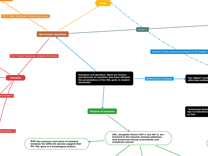
Evolution and Genetics: What are factors and theories of evolution that have affected the presentation of the VHL gene in modern mammals?
Patterns of evolution
VHL, alongside factors HIF-1 and HIF-2, are involved in the hypoxia-sensing pathways, well-preserved among invertebrate and vertebrate species
Both the presence and levels of similarly between the different species suggest that the VHL gene in a homologous feature
Duplication of ancestral hypoxia-inducible factor (HIF)α
Creation of (negative regulator) pVHL
Created the secondary contact between HIF1α Met561 and VHL Phe91 sequences
Core proteins critical for oxygen-sensing)
Possess only one HIFα gene due to substitution of VHL Phe91 with Tyr genes
Larger evolutionary divergence due to lower VHL affinity
Example: HIFα Metn-3
Primary gene present in sequence analysis of metazoan oxygen-sensing species
Present in the last common ancestor to lophotrochozoa and ecdysozoa species
Diverged in the ecdysozoa
HIFα has undergone multiple duplication events coinciding with the evolution of vertebrate species leading to great variation
Humans have three HIFα proteins
HIF1α
Most tightly bonded to VHL
HIF2α
HIF3α
"Variations in the VHL gene within different species is a result of divergent evolution, triggered by animals first diversifying between 600 and 500 million years ago under conditions that today would be described as hypoxic"
Substitutions resulted in functional divergence
Specialized hypoxic signalling relative to vertebrate needs/oxygen consumption
VHL affinity to HIFa genes
HIFα-VHL complex stability varies between species and drives adaptation to their environments
Example: HIF1α Metn-3 is the most stable HIFα-VHL complex, present in many animals
Significant variation is tolerated due to unique selective pressures
Combinations of VHL Phen+3 and HIF1α Metn-3 emerged during evolution through multiple lineages
Emerged approximately 500 million years ago in the modern-day lampreys' ancestors
Non-Human Organisms
Primates
Chimpanzees
106 bp minimal promoter region: 99% similarity
Entire VHL 5‘ sequence: 98% similarity
Diverged from humans last
Gorillas
106 bp minimal promoter region: 97% similarity
Entire VHL 5‘ sequence: 96% similarity
Macaque
106 bp minimal promoter region: 50% similarity
Entire VHL 5‘ sequence: 45% similarity
Diverged from primates earlier
Olive Baboon
106 bp minimal promoter region: 95% similarity
Entire VHL 5‘ sequence: 93% similarity
Murinae Subfamily
Mice and Rats
Four evolutionarily conserved regions were identified; Nucleotide identity similarity above 65%
Region 1: Between nucleotides +2 to +17
Region 2: Between nucleotides −49 to −19
Sequence conservation over 100 million years of evolution highlights evolutionarily conserved regions; When removed, testing showed a reduction in function of the VHL gene
Dogs
Renal cell carcinoma (RCC)
Lower prevalence of VHL mutations
Dog oncogenesis differs
90% similarity to humans
Von Hippel–Lindau: Genetic Disease affecting multiple organ systems
Presentation in humans
Genotype-Phenotype correlation
Type 1
Genetic Hallmarks
Tumors
Cysts
Type 2
Type 2A - Low risk for renal cell carcinoma
Type 2B - High risk for renal cell carcinoma
Type 2C - No risk for renal cell carcinoma
However, each variation is associated with different increased probability of presenting certain tumors/cysts depending on depending which VHL type, which cell type, and the second mutation location
Discovery
Eugen von Hippel
1904: First described rare disorder in the retina
1911: Named the disease "angiomatosis retinae"
Arvid Lindau
1923-1926: Got his PhD studying CNS tumor and cyst pathology
Named von Hippel Lindau in 1960
VHL gene was identified in 1993
"Autosomal dominant cancer-predisposition due to inheritance of a single mutated allele of VHL"
Mutation
Mutations in the nucleotides CC→ AA, in the range of 3p-21 to 3p-25
Most common genetic changes
Deletions
Deletion and certain missense mutations result in an increased risk for hemangioblastoma and RCC formation
In exons
In-frame
Frameshift
Insertions
In-frame
Frameshift
Point mutation
Most destructive in the Sp 1 region
Truncating
Missense
Associated with worse prognoses/higher rates of fatal cysts and tumors
Splice-site
Structural variations
Other gene fusions
Caused by loss of function mutations; Deletions or Truncations
Caused by missense or deletion mutation
Mutations in both copies of the VHL gene must appear for VHL to present
Chromosome 3: Controls cell growth and cell death
First allele mutation
De novo genetic change
Appears during the formation of gametes or in early stages of the zygote
1600 different pathogenic variations and over 1800 entries for genetic variation in VHL gene collected on the VHL databases
Chuvash (congenital) Polycythemia
An autosomal recessive, inheritance of germline mutations in both VHL alleles
Missense homozygous mutation of 598C>T
May result from compound heterozygous genetic changes
P192A and L188V mutations in one allele, “polycythemia-causing” p.R200W in the second allele
C-terminal region, Elongin-C binding region, or close to it
Sexual Selection: Originally isolated within the Chuvash population of Russia
Caused by a high allele frequency of VHL in a reproductively isolated area
Has since presented worldwide
Inheritance

Subtopic
Manifestation and Inheritance
Some who present similar symptoms do not have the mutation
Some with the mutation do not present with the disease
Inherited germline genetic variant
Second mutated allele
Undergoes somatic change resulting in loss of second allele
Generally unknown mutation triggers
Chance errors in cell replication
Environmental
Chemical exposure
Physical factors
VHL may skip generations; Not all people get the second mutation
50% of cases have only one mutated allele
Passed from parent to offspring
VHL is seen in all areas of the world, relatively equally in both sexes and all races
