Starch Digestion
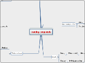
Systemic Tissues
Lipoprotein Lipase, found on the cell surface, removes fatty acids from TAG, DAG, and MAG to allow the fatty acids to enter the cell.
Fatty acids enter the cell's mitochondria to undergo Beta-Oxidation. This provides energy to the cell.
Fatty acids are stored.
Liver
HDL leaves the liver to pick up cholesterol from tissues
After HDL has picked up cholesterol and is "full", it returns to the liver.
Fatty Acids are packed up into VLDL
VLDL delivers fatty acids to tissues
When the VLDL has delivered enough fatty acids to tissues, it becomes IDL
IDL delivers fatty acids to tissues. When the IDL has delivered enough fatty acids to tissues it becomes LDL
LDL has delivered most of its fatty acids to tissues, it is mostly made up of cholesterol.
LDL delivers cholesterol to and in between cells before returning to the liver.
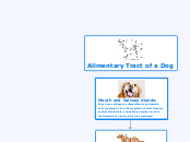
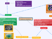
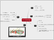
These diagrams show the Alimentary System of a Dog, the digestion of lipids, the digestion of proteins, and the digestion of starches.
The Digestion of Lipids, Proteins, and Starches in a Dog
Protein Digestion
Stomach:
The stomach is the site of the physical digestion of food.
Duodenum
Pancreatic lipase and pancreatic amylase break down fats into TAGs, DAGs, MAGs, Free Fatty Acids, Cholesterol, and Fat-soluble vitamins
Micelles of the duodenum transport the fatty acids to the enterocytes
Fatty acids are absorbed into the enterocytes of the small intestine
Long chain fatty acids are transported to the lymphatic system
Long chain fatty acids are transported through the lymphatic system via Chylomicrons, delivering fatty acids to tissues
Chylomicrons travel to the Liver
Short and medium chain fatty acids are transported to the Portal Vein
Short and medium chain fatty acids enter the Liver
Bile emulsifies fats to make room for pancreatic lipase
Alimentary Tract of a Dog
Duodenum:
The duodenum is the first section of the small intestine. The duodenum receives pancreatic secretions from the pancreatic duct. Non-enzymatic digestion in the duodenum includes the emulsification of fats (by bile) and Cholecystokinin hormone. The duodenum is also home to many zymogens needed for starch and protein digestion.
Jejunum:
The jejunum is the second section of the small intestine. It has the most surface area of the small intestine. Most nutrient absorption occurs in the jejunum.
Ileum:
The role of the ileum, the third section of the small intestine, is residual nutrient absorption.
Cecum:
The cecum is the first section of the large intestine. The cecum is a blind pouch at the junction of the small and large intestines. It's role includes microbial digestion of fibrous carbohydrates, microbial production of volatile fatty acids, and the synthesis of B-Vitamins.
Colon:
The colon is the second section of the large intestine. It functions in microbial digestion of fibrous carbohydrates, microbial volatile fatty acid synthesis, synthesis of B-vitamins and Vitamin K, and water reabsorption and concentration of feces.
Rectum:
The rectum is the final section of the large intestine where undigested material (such as unabsorbed or undigested feedstuffs, dead bacteria, sloughed cells, and fluid) is formed into feces.
Anus:
The anus, the last part of the alimentary tract, controls the exit of feces.
Small Intestine consists of:
Duodenum, Jejunum, Ileum
Duodenum:
Pancreatic Amylase of the duodenum, continues to hydrolyze the starches into monosaccharides. Maltase, sucrase, and lactase digest their respective sugar molecules.
Any remaining, undigested starches in the small intestine (duodenum, jejunum, or ileum) are hydrolyzed to monosaccharides by brush border enzymes.
Monosaccharides are absorbed by the enterocytes of the small intestine and into the blood stream.
Monosaccharides arrive at the liver, via the portal vein.
If in excess, glucose is converted to acetate to make triglycerides (fat).
Glucose is stored as glycogen, for future energy.
Glucose (a monosaccharide) is absorbed into the cells by insulin to provide energy.
Mouth and Salivary Glands:
Salivary amylase begins to catalyze the hydrolysis (breakdown) of starches.
Esophagus
Stomach:
The stomach is the site of the physical digestion of starches.
Duodenum:
Enzymes in the Duodenum that digest polypeptides (protein):
Trypsinogen
Chymotrypsinogen
Proelastase
Procarboxypeptidase A
Procarboxypeptidase B
These enzymes continue to hydrolyze (break) peptide bonds until there are no peptide bonds left. The result is free amino acids.
Amino acids are then absorbed by enterocytes of the small intestine to the bloodstream.
Amino acids arrive to the liver via the portal vein.
The carbon skeleton of proteins can be used for ketogenic or glycogenic energy.
Amino acids are used in enzyme and hormone synthesis.
Amino acids are used for tissue protein synthesis.
Mouth and Salivary Glands
Esophagus:
Stomach:
The stomach is the site of the chemical digestion of food. Chief cells of the stomach produce HCl, which is needed to activate pepsinogen into pepsin. Pepsin is the active enzyme which digests protein by breaking peptide bonds, forming polypeptides.
Lipid Digestion
Mouth and Salivary Glands:
Dogs have sublingual, submandibular, and parotid salivary glands. The salivary glands consist of water, sodium bicarbonate, and salivary amylase to aid in the formation of a bolus, which is swallowed.
Esophagus:
The esophagus is a muscular tube of circular and longitudinal smooth muscle. The esophagus is needed for swallowing. Food is pushed down the esophagus by peristalsis, or muscular contractions that push feed to the next organ.
Stomach:
The stomach is the site of the chemical and physical digestion of protein. In the stomach HCl, produced by chief cells, activates pepsin (important enzyme for protein metabolism).
