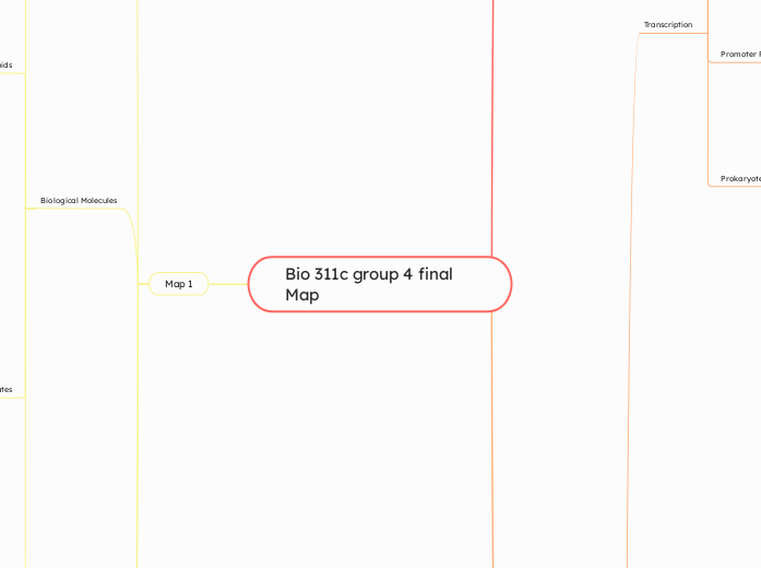
Bio 311c group 4 final Map
Map 1
Cellular Functions and Organelles
Animal Only
Extracellular Matrix
Collagen
Fibronectin
Protoglycan
Integrins
Tight Junction
Desmosome
Gap Junction
Present in both Animal and Plant Cells
Mitochondria
Vesicles
Nucleus
Nuclear envelope
vacuoles
Endoplasmic Reticulum
Rough ER
Ribosomes
Smooth ER
Golgi Apparatus
Lysosome
Peroxisome
Cytoskeleton
Microtubules
Microfilaments
Intermediate Filaments
Plant Only
Plasmodesmata
Cell wall
Central Vacuole
Chloroplast
Prokaryotic Only
Biological Molecules
Protiens
Amino Acid
Main Chain
Amino Group
Carboxyl Group
Side Chain (R-Group)
Polar
Nonpolar
Basic
Acidic
Protien Folding
Primary
Peptide Bonds
Dehydration Synthesis
Secondary
Hydrogen Bonds
Types
Alpha Helices
Beta Pleated Sheets
Tertiary
Interaction of R-Group
Hydrogen Bonds
Ionic Bonds
Van Der Waals
Disulfide Bonds
Quaternary
Interchain Interactions
Protien Structure
Amino Acid Sequence
Physical/Chemical Conditions
pH
Temperature
Salt
Solution Prevents Disulfide Bond
Lipids
Fat Molecule
Glycerol
Fatty Acids
Unsaturated
Liquid at Room Temperature
Isomers
Trans
Cis
Double Bond
Saturated
Solid at Room Temperature
Uses Ester Linkage
Dehydration Synthesis
Phospholipids
Form Closed Liquid Bilayers
Parts
Hydrophilic Head
Hydrophobic Tails
Steroids
Cholesterol
Amphipathic
In Cell Membrane
Types
HDL
LDL
Carbohydrates
Polysaccharides
Structure
Cellulose
Linear
No Branching
Storage
Glycogen
Extensive Branching
Starch
Amylose
No Branching
Amylopectan
Some Branching
Use Glycosidic Linkage
Alpha 1, 4
Alpha 1, 6
Beta 1, 4
Bet 1, 6
Glucose
Alpha
Glycogen
Starch
Beta
Cellulose
Nucleic Acids
Types
DNA
Ribose
Thymine
Double-Stranded
Complementary Base Pairing
Uses Hydrogen Bond
RNA
Deoxyribose
Uracil
Single-Stranded
Both
Base Pairing
Use Hydrogen Bonds
Sugar-phosphate Backbone
Nucleotides
Deoxyribose Sugar
Phosphate Group
Nitrogenous Base
Cytosine
Thymine
Uracil
Adenine
Guanine
Use Phosphodiester Bond
Dehydration Synthesis
Nucleosides
Deoxyribose
Nitrogenous Base
Chemical Bonds
Intermolecular Forces
Van Der Waals
Hydrogen Bonding
Hydrophobic
Dipole-Dipole
Ion-Dipole
Intramolecular Forces
Covalent
Non Polar Covalent
Polar Covalent
Ionic
Bond Strength
Electronegativity
Water
Water Properties
Cohesion
Water Transport in Plants
Surface Tension
Adhesion
High Specific Heat
Helps moderate temperature
High Heat of Vaporization
Evaporative Cooling
Expansion Upon Freezing
Denser a Liquid than a Solid
Universal Solvent
Acids and Bases
pH
Acidic
Basic
Neutral
pOH
Molecules interacting with water
Hydrophobic
Hydrophilic
Amphipathic
Molecular Structure
Bent
Polar
Bonds
Hydrogen Bonding
Water molecules & heat
Heat is absorbed
Hydrogen bonds break
Heat is released
Hydrogen bonds form
Map 2
Cellular Respiration
Aereobic(requires Oxygen)
Glycolysis
Takes Place in Cytosol
Inputs: 1 Glucose, 2 ATP
Outputs
Net: 2 Pyruvate, 2 NADH, 2 ATP
Total: 2 Pyruvate, 2 NADH, 4 ATP
Uses Substrate level Phosphorylation to produce ATP
Step 1: Hexokinase converts Glucose to Glucose 6 Phosphate
Step 3: Phosphofructokinase converts Fructose 6 Phosphate to Fructose 1,6 Biphosphate
Pyruvate Oxidation
Inputs: 2 pyruvate, 2 CoA
Outputs: 2 Acetyl CoA , 2 NADH
Takes place in Cytosol then mitochondrial matrix
krebs cycle
Takes place in mitochondrial matrix
Inputs: 2 Acetyl CoA
Outputs: 6 NADH, 2 FADH2, 2 ATP
Uses Substrate level Phosphorylation to produce ATP
Step 1: Acetyl CoA adds its 2 Carbon groups to Oxaloacetate, forming Citrate
Step 3: Isocitrate is oxidized to alpha ketoglutarate while NAD+ is reduced to NADH
Oxidative Phosphorylation
Takes place in inter membrane space
Inputs: O2, 10 NADH, 2 FADH2
Outputs: H2O, 26-28 ATP
Uses Substrate level Phosphorylation to produce ATP
Paired Process
Electronic Transport Chain
As electrons are transferred down ETC, the energy released is used to pump H+ against concentration gradient
Chemiosmosis
H+ transported down concentration gradient using ATP Synthase (facilitated diffusion), produces lots of energy which is used for Pi+ADP=ATP
Anaerobic(doesn't require O2)
Glycolysis (info in aerobic section)
fermentation
Alchohol fermentation
inputs: 2 Pyruvate, NADH
outputs: ethanol, NAD+
Lactic Acid Fermentation
Inputs: 2 Pyruvate, NADH
Outputs: Lactate, NAD+
Takes Place in Cytosol
Glucose oxidized, Oxygen Reduced
Energy Transfer
Metabolic Pathways
Catabolic Pathways
Cellular Respiration
Anabolic Pathways
Biosynthetic Pathways
Polymerization
Photosynthesis
Thermodynamics
System
Closed System
Open Sytem
Surroundings
Laws
1 - Energy can be transferred, but not created/destroyed
2 - Energy transfer increases entropy
Energy Changes
Exergonic (ΔG0)
No Change (ΔG=0)
Energy Coupler
Powered by ATP
Transport
Mechanical
ATP Cycle
Enzymes
Catalytic Cycle
Binding of Substrate
Lowers Activation Energy
Enzyme Activity
Temperature
pH
Substrate Concentration
Function
Normal Binding
Competitive Inhibition
Noncompetitive Inhibition
Allosteric Regulation
Activators
Inhibitors
Cooperativity
Feedback Inhibition
Communication and Signaling
Cell communication
Physical Contact
Gap junctions
Plasmodesmata
Cell surface proteins
Types of signaling
Local signaling
Long distance
Cell signaling
Reception
Membrane receptors
Hydrophilic signal
G protein linked receptor
Signal binds to GPCR causing a change in GCPR shape allowing G protein to bind to it. GDP is replaced with GTP on the G protein. The activated G protein can now activate a nearby enzyme.
Ion channel receptor
Signal molecule binds to the receptor and the gate allows specific ions through a channel in the receptor
Intracellular receptors
Steroid hormone receptor
Transduction
cAMP
Binds and activates protein kinase which goes on to activates other kinases
Phosphorylation Cascade
Kinases are activated by the addition of a phosphate group
Enzymes
Kinase
Catalyze the transfer of phosphate groups from ATP to proteins
Phosphotase
Removes a phosphate group from proteins
Adenylyl Cyclase
Converts ATP to cAMP
Phosphodiesterase
Converts cAMP to AMP
Cell Membranes
Structure and Function
Phospholipid Bilayer
Hydrophilic Head
Phosphate and Glycerol
Hydrophobic Tail
Fatty Acids
Membrane Proteins
Integral (Embedded)
Peripheral (Surface Attached)
Selective Permeability
Small Nonpolar Molecules (O₂, CO₂)
Pass Freely
Large and Polar
Require Transport
Membrane Fluidity
Cholesterol
Temperature
Saturated Fatty Acids
Unsaturated Fatty Acids
Membrane Proteins and R-Groups Orientation
Hydrophilic R-Groups
Face Aqueous Exterior/Interior
Hydrophobic R-Groups
Embedded in Lipid Bilayer
Protein Structure Bonds
Primary
Peptide Bonds
Secondary
Hydrogen Bonds
α-Helices
β-Sheets
Tertiary
Disulfide Bridges
Ionic
Hydrophobic Interactions
Quaternary
Multiple Polypeptides Interacting
Map 3
Subtopic
DNA structure and replication
DNA Structure
Phosphodiester bond to link DNA and RNA monomers
Double Helix with two anti parallel strands
Nucleotide
Nitrogenous base
Phosphate group
Deoxyribose sugar (ribose sugar for RNA)r
Subtopic
Experiments demonstrating DNA is the genetic Material
Griffith’s Transformation Experiment (1928)
Setup: Streptococcus pneumoniae strains in mice -Smooth (S) strain: virulent, capsule‑producing -Rough (R) strain: non‑virulent, no capsule
Results: Live S → mice die
Live R → mice live
Heat‑killed S → mice live
Live R + heat‑killed S → mice die; live S cells recovered
Conclusion: Genes Transferable between Bacteria
Hershey-Chase Blender Experiment
Labeling: Phage protein with 35S (sulfur in protein)
Phage DNA with 32P (phosphate in DNA):
Procedure: allow phage to infect bacteria and blend
Observations after blending: 32P (DNA) found inside bacterial pellet
35S (protein) remained in the supernatant
Conclusion: DNA is what carries the genetic information not Protein
Chargaff's rule
In any double‑stranded DNA:
AA = TT and GG = CC (base‑pair stoichiometry)
Total purines (A+G) = total pyrimidines (C+T)
DNA Replication
Meselson-Stahl Experiment
Labeling: Grow E. coli in 15N (“heavy” nitrogen) then shift to 14N (“light”) medium.
Results after Centrifugation: Gen 1 → single intermediate band
Gen 2 → intermediate + light bands
Conclusion: Semiconservative Replication
3 types of replication:
Conservative: Original double helix remains intact; a wholly new duplex is synthesized.
Semiconservative: Each daughter duplex contains one parental strand and one new strand.
Dispersive: Both strands of both daughter duplexes are hybrids of old and new segments.
Parts of replication:
Origin of replication(ORI): Specific DNA region where replication begins (AT‑rich for easier strand separation)
Replication bubble: Local unwound region around ORI
Replication forks: The two Y‑shaped junctions at bubble edges where new strands grow
Enzymes for Replication
Leading-Strand Synthesis Enzymes
Helicase: unwinds the DNA duplex
Single‑strand binding proteins (SSBs): stabilize unwound strands
Primase (RNA polymerase): lays down short RNA primer
DNA polymerase III: extends primer continuously
Lagging Stand Synthesis (Okzaki Fragments)
Primase: synthesizes multiple RNA primers at intervals
DNA polymerase III: extends each primer to make Okazaki fragments
DNA polymerase I: removes RNA primers and replaces with DNA
DNA ligase: seals nicks by forming phosphodiester bonds
Bidirectional & Discontinuous Replication
Bidirectional: Two replication forks move in opposite directions from ORI.
Continuous (leading) strand: Synthesized 5′→3′ in the direction of fork movement.
Discontinuous (lagging) strand: Synthesized as short 5′→3′ Okazaki fragments away from fork; later joined.
Transcription
3 steps
Initiation
Elongation
Termination
Visual Interpretation
Start Site: +1
Upstream: -1, -2
Downstream: +2, +3
written 5' to 3'
Uracil(U) instead of Thymine(T)
Template strand 3' to 5'
Promoter Region
where RNA Polymerase and necessary transcription factors bind
Prokaryote Vs. Eukaryote
Prokaryotes
Location: Cytoplasm
RNA polymerase used
mrna, no pre-mrna
coupled w/ translation
Eukaryotes
Location: Nucleus
RNA polymerase 2
has initial pre-mrna
Poly A site, Poly A tail placed by Poly A Polymerase
5' Cap
promoter has TATA box
Transcription factors
Spliceosomes cleave introns out of Pre-mrna
Alternate splicing: Remove some introns, keep others , results in one gene expressing diverse rpteins
Gene Regulation
Prokaryotes
Operon
Promoter
Operator
Positive Regulation
Activator = On
No Activator = Off
Negative Regulation
Repressor = Off
No Repressor = On
Structural Genes
Lac Operon Example
Regulatory Sequences
Regulatory Gene
LacI
Promoter for Regulatory Gene
Promoter for Structural Genes
Operator
Lac Operon
Structural Genes
LacZ (B-galactosidase)
LacY (Permease)
LacA (Trans-acetylase)
Operator
Promoter
Options
No Lactose
Active Repressor
Operon = Off
Has Lactose
No Glucose
High Camp Level
Operon = On
Inavtive Repressor
Operon = On
Inavtive Repressor
Operon = On
Has Glucose
Low Camp Level
Operon = Off
Eukaryotes
Nucleosomes
Histone Protiens
H1
H2A
H2B
H3
H4
DNA
Histone Core
H2A
H2B
H3
H4
Steps
10-nm Fiber
DNA winds around histones
30-nm Fiber
fiber coils/folds
300-nm Fiber
forms looped domains
Metaphase Chromosome
coil further
Differential Gene Expression
Transcription Initiation
Transcription Factors
General
Basal
Specific
Activators
Repressors
Control Elements
Proximal
Sequences near promoter
Bind general transcription factors
Distal
Sequences upstream/downstream
Enhancers
bind specific transcription factors
Process
Acivator binds to enhancer
DNA bending protien brings activators to promoter
Activators bind to mediator protiens
Liver Cell vs Lens Cell Example
Lens Cell
Albumin Gene = Not Expressed
Crystallin Gene = Expressed
Liver Cell
Albumin Gene = Expressed
Crystallin Gene = Not Expressed
Translation
Accurate translation
Correct match between tRNA codon and mRNA codon
Aminoacyl-tRNA synthase
Correct match between tRNA and amino acid
Process
Initiation
Prokaryotes
Translation initiation complex
mRNA, tRNA carrying 1st amino acid, and 2 ribosomal subunits are brought together
Requires GTP
1st amino acid
Formyl-methionine
Eukaryotes
Small ribosomal subunit and tRNA first bind the 5' cap
Scans mRNA to find first start codon (AUG)
Then large ribosomal subunit comes to form translation initiation complex
1st amino acid
Methionine
Elongation
3 binding areas
P site
Holds the tRNA that carries the growing polypeptide chain
A site
Holds the tRNA that carries the next amino acid
E site
Exit site where tRNA leave the ribosome
Starts when tRNA carrying next amino acid comes to A site
Peptidyl transferase forms peptide bond between the 2 amino acids
tRNA in P site is now empty and moved to E site to be released
tRNA from A site moves to P site
New tRNA comes to A site
mRNA read in 5' to 3' direction
Amino acids added in N to C direction
Termination
Stop codon is reached in A site
Release factor sits in A site disassociating the translation initiation complex
GTP driven
Components of translation
tRNA
Single RNA strand about 80 nucleotides long
Bring correct amino acid to be added
Ribosomes
Prokaryotes
70S ribosomes
Eukaryotes
80S ribosomes
Large and small ribosomal subunit
Made of proteins and ribosomal RNA's
Codon
Set of 3 amino acids corresponding to an amino acid
Codon table
Universal
Degenerate
Noneverlapping
