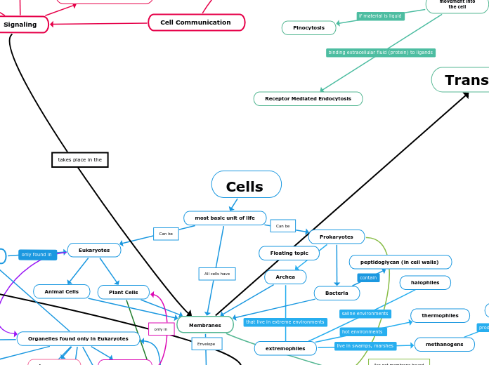
Cells
most basic unit of life
Eukaryotes
Animal Cells
Plant Cells
Photosynthesis
Light-dependent Reactions
Thylakoid Membrane
Chloroplasts
Chlorophyll
Natural Pigment
Water
Energy from Sunlight
NADPH
ATP
O2
Calvin Cycle
Stroma
ATP
NADP
NADPH
Glucose
ADP + P
C4 Photosynthesis
PEP Carboxylase
Oxaloacetate
Malate
Bundle Sheath Cells
CO2 + Pyruvate
CAM Photosynthesis
PEP Carboxylase
Oxaloacetate
Malic Acid
Vacuole
takes place at night
H20 loss
Light Reactions
Photosystem II P680
Energy
Prokaryotes
Bacteria
peptidoglycan (in cell walls)
Archea
extremophiles
halophiles
thermophiles
methanogens
methane as waste product
Cytoplasm
Organelles
Organelles found only in Eukaryotes
Nucleus
Contains genetic material
Mitochondria
ATP
evolved from bacteria
Golgi apparatus
Storage and Transport
Vacuoles
Storage
Lysosomes
Breaks down cell waste
Rough Er
Storage for Protien
Smooth ER
Storage for protein
and lipids
Chloroplast
Produces Sugar
Found in Eukaryotic and Prokaryotic cells
Ribosomes
Protein Synthesis
DNA
RNA
Uracil
Thymine
mRNA
Codon
Start Codon
Stop Codon
tRNA
Anticodon
Amino Acids
Ribosomes
A site
B site
Exit Site
mRNA + tRNA
tRNA on P-site
moves along mRNA making polypeptide chain
Stop Codon
Protein is Released
all parts detach for each other
Point Mutations
Base Pair Substitution
Nonsense
premature stop codon is present
Silent
there is no effect on amino acid produced
Missense
wrong amino acid is produced
Insertions and Deletions
Frameshift Mutations
Immediate Nonsense
Extensive Missense
Missing/Extra Amino Acid
Rough ER
Glycosylated
Golgi Complex
Flagella
Located on the outside of the cell
Allows the cell to move
Prokaryotic Organelles
Are not membrane bound
Pili
Motion, attaching to objects,
transfer of genetic material
Nucleoid
genetic material, is not membrane bound
Transport
energy
smalls amounts of substances
pass through proteins (pumps)
Sodium Potassium Pump
3Na+ kicked out against
concentration gradient
2K+ are brought in against
concentration gradient
conformation/changes in shape
large amounts of substances
pass through membrane (vesicles)
Exocytosis
movement out
of the cell
Endocytosis
movement into
the cell
Phagocytosis
Pinocytosis
Receptor Mediated Endocytosis
no energy
Simple Diffusion
Facilitated Diffusion
substances cannot cross
membrane on their own
Hydrophilic Molecules
Channel Protein
uses channel for molecules
and specific solutes
Hydrophobic Molecules
Carrier Protein
shapeshifts to help
different molecules
Osmosis
water crosses the
the cell membrane
Hypotonic
Turgor
Isotonic
Normal
Flaccid
Hypertonic
Shriveled
Plasmolyzed
contractile vacuole
taking water in
pumping water out
movement of substances
in and out of the cell
Phospholipid Bilayer
Amphipathic
Two Hydrophobic Tails
Saturated Fatty Acid
no double bonds
between carbons
Unsaturated Fatty Acid
double bonds
between carbons
Trans Fatty Acids
H atoms are opposite
Cis Fatty Acids
H atoms are on
same sides
Hydrophilic Head
Phosphate Group
Glycerol
Lipids
Cholesterol
Carbohydrates
Glycolipid
Protein
Glycoprotein
Peripheral Membrane
Proteins
Integral Membrane
Proteins
All cells have
Can be
Can be
Biological Molecules
Include
Carbohydrates
Consisting of 1 Sugars
Monosaccharide
Fructose
Glucose
composed of alpha and beta isomers
Consisting of 2 Sugars
Disaccharide
Examples
Sucrose
Lactose
formed when a dehydration reaction joins two monosaccharides
Consisting of 2 or More Sugars
Polysaccharide
Examples
Glycogen
Animals
Used For
Storage of Energy
Formed of Alpha Glucose monomers connected through 1-4 glycosidic linkages
Starch
Can be Found Within
Plants
Proteins
Are Chains of
Amino Acids
Polypeptides
Examples
Enzymes
Hemoglobin
Can Be
Primary
peptide bonds
Tertiary
Subtopic
polar covalent, ionic, hydrophobic interactions, and R-group disulfide bonds
Quaternary
several protein chains or subunits into a closely packed arrangement
all prior bonds, van der waals
3 Main Groups
Fat (Triglycerides)
Unsaturated
Presence of double bonds can create cis/trans isomers of fatty acids
stay liquid at room temperature
cis isomer
H atoms on same side of molecule
trans isomer
H atoms on opposite sides of molecule
Saturated
only single bonds between neighboring carbons in the hydrocarbon chain, saturated with hydrogen
are solid at room temperature
Phospholipid
the structural components of the cell membrane (phospholipid bilayer)
hydrophillic head, 2 hydrophobic tails
“kinks” in their tails push adjacent phospholipid molecules away, which helps maintain fluidity in the membrane
Steroids
serve as structural components of cell membranes, as energy storehouses, and as important signaling molecules
Nucleic Acids
Nucleotides
Phosphate Group
Adenine
Guanine
Thymine
Cytosine
Uracil
ATP
includes phosphodiester bonds
5 Carbon Sugar
Ribose
RNA
Deoxyribose
DNA
nucleic acids
carries genetic information
synthesize proteins
replicates itself
Purines
Adenine (pairs with Thymine in DNA, Uracil in RNA) and Guanine (pairs with Cytosine)
A-T form two hydrogen bonds C-G forms 3 hydrogen bonds
The amount of Adenine equals the amount of Thymine, the amount of Guanine equals the amount of Cytosine
Pyrimidines
Thymine (pairs with Adenine in DNA, Uracil (pairs with Adenine in RNA), and Cytosine (pairs with Guanine)
DNA Replication
semi-conservative model of replication
The two strands of the parental molecule separate, and each functions as a template for synthesis of a new, complementary strand
helicase
is an enzyme that catalyzes DNA strand separation
breaking hydrogen bonds between strands
creating a replication fork/bubble for replication to begin
primase
binds to each strand of DNA at the replication fork and synthesizes a short (3 to 10 base) strand of RNA
primer
provides a strand end for DNA polymerase to add bases to
DNA polymerase III
adds complementary nitrogenous base to daughter strand
lagging strand
numerous RNA primers, made by the primase enzyme, bind at various points along the lagging strand
chunks of DNA are then added to the lagging strand also in the 5’ to 3’ direction
Okazaki fragments
leading strand
binds to the leading strand and adds new complementary nucleotide bases (A, C, G and T) to the strand of DNA in the 5’ to 3’ direction
DNA Polymerase I
removes the RNA primer and replaces with DNA nucleotides
DNA ligase
seals up the sequence of DNA into two continuous double strands
sliding clamp protein
converts the DNA polymerase III from being distributive (falling off) to processive (staying on)
SSB (single strand binding proteins)
stabilize the un-wound parental strands
topoisomerase
alters the supercoiling of double-stranded DNA
dna synthesis
Example
DNA, RNA
Bonds
covalent
polar
unequal sharing of electrons
O-H, S-H, N-H, C-O
hydrophillic
equal sharing of electrons
C-H, C-C, H-H
hydrophobic
salts, ex. NaCl
ionic bonds
Envelope
Surround
Is attached to the
only in
Secondary
hydrogen bonding
relatively weak
relatively strong
sharing of H atom
partial positive charges that attract partial negative charges
among H2O molecules
water properties
cohesion
water molecules attracted to other substances
adhesion
water molecules attracted to each other
ex. water move up stem of a plant
high specific heat
amount of heat needed to raise the temperature of 1 gram of a substance 1 degree Celsius
water has highest specific heat of an liquid
hydrogen bonds breaking and reforming
high heat of vaporization
amount of energy to convert 1g or a substance from a liquid to a gas
hydrogen bonds to be broken
water expansion when frozen, denser as liquid than solid
ice has spaced-out lattice structure
ice to float
aquatic life can survive under frozen water
universal solvent
water is a polar molecule
Can be attached to
Provides
Provides
amino acid chains and polypeptide backbone held together by
alpha helices, beta pleated sheets
Floating topic
formed when 100 or more monosaccharides are bonded together through glycosidic linkages
Function:
Photosynthesis
Cellular Respiration
three distinct processes
Glycolysis
cytoplasm
Energy Investment Phase
step 1- adding a phosphate from ATP to Glucose to form Glucose6p by using the enzyme Hexokinase
step 2- converting Glucose6p into Fructose6p
step 4- 6 carbon sugar splits into two molecules of 3 carbon. Each of them forming DHAP and G3P
step 5- conversions between the DHAP
Energy Payoff Phase
step 6- 1 G3P is oxidized by the transfer of electrons to NAD+, and forming NADH. Using energy from the redox reaction, a phosphate group, is then attached to the oxidized substrate, making 3-Biphosphoglycerate.
step 7- the phosphate group is transferred to ADP in an exergonic reaction.G3P is then oxidized to the carboxyl group, creating 3-Phosphoglycerate
step 9- Enolase causes a double bond to form in the substrate by extracting a water molecule, creating phosphoenolypyruvate (PEP)
breaks down glucose into two molecules of pyruvate
Lactic Acid Fermentation
NADH transfers its electrons directly to pyruvate
glucose is converted to lactate and cellular energy
animal cell tissue
yeast, muscle cells, and Lactobaccilus ssps
occurs in production of cheese and yogurt
pyruvate decarboxylase and lactate dehydrogenase
Alcohol Fermentation
NADH donates its electrons to a derivative of pyruvate
carboxyl group is removed from pyruvate and released in as carbon dioxide
a two-carbon molecule called acetaldehyde
regenerating NAD+ and forming ethanol
pyruvate decarboxylase and alcohol dehydrogenase
plant tissue, yeast, microorganisms
occurs in production of bread, beer, wine, vinegar
Pyruvate Oxidation
Acetyl Coenzyme A is formed
Oxidative Phosphorylation
Electron Transport Chain
located in the inter mitochondrial membrane
Complexes I, III, IV
are H+ pumps
their job
pump H+ against the concentration gradient in the intermembrane space
the energy used coms from the energy released from the electrons being transferred down the ETC
Complex II
FADH2 transfers electrons
an electrochemical gradient that leads to the creation of ATP
Creates ATP
H+ go back down the concentration gradient
ATP synthesis
Citric Acid Cycle
step 1- Acetyl CoA adds its two carbon group to oxoloacetate which produces citrate
step 2- Citrate converts to Isocitrate, with the loss and gain of an H2O molecule
step 3- * Isocitrate is oxidized while NAD+ is reduced. a-Ketoplutarate is made
step 4- CO2 is released from a- ketoglutarate, which causes four-carbon molecules to be oxidized, then CoA is added, which makes it reactive, NADH+ & H+ is formed. CoA-SH is added, creating Succinyl CoA
step 5- A phosphate group is added to Succinyl CoA. GTP is released, which binds with ADP, that results in the formation of ATP. COA-SH is released, and Succinate is the end result.
step 6- Succinate is oxidized, FAD is reduced, FADH2 is formed
step 7- H2O is added to Furmarate, which results in Malate
step 8- Malate is oxidized, NAD+ is added, and it results in the formation of NADH+. The product is Oxaloacetate
dependent on each other as the products of each of these reactions initiate the other reaction
Signaling
Local Signaling
Synaptic Signaling
Nerve cell signaling
1) An action
potential arrives,
depolarizing
the presynaptic
membrane.
2) depolarization
opens voltage-gated
channels
Triggering an
influx of Ca2+
3) elevated Ca2+ concentration
causes synaptic vesicles to fuse with
the presynaptic membrane
neurotransmitter is released
into synaptic cleft
4) neurotransmitter binds to ligand-gated
ion channels
Paracrine Signaling
Long Distance Signaling/
Hormonal Signaling
Hormone travels through blood steam
to reach target cell
Signal Molecule/
ligand
Signal must be received
by a receptor
signal is
polar/hydrophilic
Membrane
Receptor
G Protein
coupled receptor
1) Ligand binds to
extracellular receptor
receptor changes shape
and activates G protein
2) activated G protein leaves the receptor, diffuses along the membrane, and then binds to an enzyme
enzyme changes shape
and becomes activated
3) Enzyme takes ATP
and makes cAMP(2nd
messenger)
4) cAMP activates
Protein Kinase A
Triggers cellular
response
Ion Chanel
signal molecule attaches to the
Ligand-gated ion receptor
channel opens
specific ions are allowed
through the channel into the
cytoplasm
Triggers Cellular
response
When ligand leaves
receptor, channel closes
Tyrosine Kinase
Receptor tyrosine
kinase proteins
(inactive monomers)
(2 of them)
each receptor
has 3 tyrosines
1) 2 Signaling molecules
bind to the 2 binding sites
the monomers combine
to create a dimer
2) 6 ATP is used to
add a Pi to each tyrosine
activating the tyrosine
kinase dimer
2) Inactive relay proteins
attach to the tyrosine dimer
relay proteins become
activated
Triggers Cellular
response
Signal is non-polar
hydrophobic
Intermembrane
Receptor
1) steroid hormone
passes through the
plasma membrane.
2) hormone binds
with receptor protein
and activates it
3) hormone-receptor complex
enters the nucleus and binds
to specific genes.
bound protein acts
as a transcription factor
4) stimulating the
transcription of
the gene into mRNA
5) mRNA is
translated into a
specific protein.
takes place in the
Cell Communication
Physical Contact
Junctions
Gap
Open channel between
cells
Allows free flow of
ions and small molecules
Tight
prevent fluid from moving
across cells.
Plasmodesmata
Chanel between cells
only found in plant cells
Desmosomes
Provides a connection
between intermediate
filaments
Somewhere between gap and tight!
cell releases
signal molecule
Photolysis
Two Electrons
Photosystem I P700
ETC
2 NADH
Primary Electron Acceptor
Metabolism
Catabolism
breaking down of molecules
releases energy
Energy of Life
Chemical Energy
ATP
Thermodynamics
Conservation of Energy
energy cannot be created/destroyed
entropy always seeks to increase over time
Anabolism
building up of molecules
requires energy
Floating topic
comprised of nucleotides made of nitrogenous bases
Transcription
mRNA
DNA
pre-mRNA
eukaryotes
stability
translation
exons
expressed
introns
spliced out
snRNP's
ribosomes
promoter
stop codon
DNA- dependent RNA polymerase
in 3' to 5' direction
DNA polymerase
in 5' to 3' direction
terms
downstream
upstream
toward 3' end of RNA
toward 5' end of DNA
Translation
mRNA
small ribosomal
subunit
tRNA carries
anti codon
Start Codon
(AUG)
Large Ribosomal
subunit joins complex
P site
A site
anti codon pairs with
corresponding codon
E site
(exit site)
tRNA in A site moves to
Codons
Sequence of 3 Nucleotides
Stop Codon
Ends translation
UAA, UAG, UGA
Start Codon
Begins Translation
AUG
Cytosol
Floating topic
Sequences
Sequence
next tRNA
enters
here
tRNA in P site
moves to
Contains
binds to
binds to
Process that creates
initiation complex
complete
Initiation Complex
part of
first tRNA
starts in
Elongation Begins
begins with
Aminoacyl tRNA synthetase
attaches corresponding
amino acid to
As tRNA travels from
A site to P site
Peptidyl transferase
Peptide bonds
Amino acids
Polypeptide chain formed
End codon is reached
Release factor
Complex breaks
apart
Both ribosomal units
Polypeptide chain
tRNA
(w/o amino acid)
