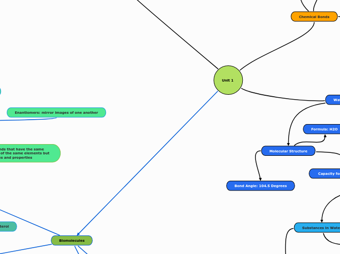
Unit 1
Biomolecules
Carbohydrates
Three Types of Isomers
Structural Isomers: differ in the covalent arrangement of their atoms
Geometric Isomers
Cis Isomer: same side
Trans Isomer: opposite side
Enantiomers: mirror images of one another
Sugars and polymers of sugars
Monosaccharides: simplest sugars; made of C, H, OH, and CO groups
Ketoses: CO group is in the middle of the chain
Aldoses: CO groups at the middle of the chain
Disaccharide: formed when a dehydration reaction joins two monosaccharides
Glycosidic Linkage: formed through covalent bond
Polysacchrides: formed when 100 or more monosaccharides are bonded together through glycosidic linkages
Storage
Dextran
Structure
Cellulose
made of beta-Glucose; no branching; insoluble fiber
Chitin
Lipids
Fats
made of glycerol and three fatty acids
compact way for animals to carry their energy stores with them
connected through ester linkage
Ester Linkages: connects each fatty acid to an OH in glycerol
can contain one tupe of different types of fatty acids
Saturated
solid at room temp
found in animal sources
no double covalent bonds between carbons; saturated with hydrogen atoms
increased incidence of cardiovascular disease
Unsaturated
come from plant sources
liquid at room temp
one or more double covalent bonds are found within carbon chain; do not have hydrogen atoms at every position
Cis: presence of double bonds; slight kinks
Trans: trans fatty acids
Steroids
four fused rings
HDL: high density lipoprotein or "good cholesterol"
LDL: low density protein or "bad cholesterol
increased by saturated fats and trans fat
Nucleic Acids
Deoxyribonuleic Acid (DNA)
provides directions for its own replication
directs synthesis of messenger RNA and control protein synthesis (gene expression)
transcription: information in the DNA is used to make mRNA
translation:information from the mRNA is used to make proteins
A, G, C, T
doubled stranded
Ribonucleic Acid (RNA)
A,G,C,U
single-stranded
polymers made of monomers Nucleotides
5 carbon sugar
phosphate
nitrogenous base
purines: A, G
pyrimidines: C, T(U in RNA)
Nucleosides
does not have a phosphate group
connected through phosphodiester bonds/linkages through condensation/dehydration reactions
Proteins
made of monomers called amino acids
has a central cabron surrounded by amino, carboxyl, hydrogen, and R group
made of main chain and side chain
four types of basic groups
Polar
contains OH, SH, or NH groups
Nonpolar
contains H, CH, or carbon ring; hydrophobic
Acidic (-)
Basic (+)
Chemical Bonds
Molecular Function
Size and Shape are key to function.
Intramolecular
Covalent Bonds
Nonpolar
Polar
Electronegativity Differences (Greater differences give more polar bonds)
Ionic Bonds
Crystalline Structures (Salts)
Intermolecular
Dipole-Dipole
Hydrogen Bonds
Strong Dipole-Dipole Interactions between H and O, F, or N (Partial Positive H with Partial Negative O, F, N).
Ion-Dipole Interactions
Hydrophobic Interactions
van der Waals Interactions
Water Properties
Properties of Water
Cohesive Behavior
High Surface Tension
Water Transport in Plants
High Specific Heat
Temperature Moderation
High Heat of Vaporization
Evaporative Cooling
Expansion Upon Freezing
Most Dense at 4 Degrees Celsius
Stable Hydrogen Bonds; Ordered
Less Dense as a Solid
Floats on Water; Insulation of Ice for Waters Below
Universal Solvent
Dissolves most substances
Substances in Water
Nonpolar Substances
Water Forms a Cage Around Nonpolar Molecule
Polar Substances
Polar and Ionic Compounds Dissolve in Water
pH
con
Bases decrease Hydronium Concentration (More OH- groups).
pH + pOH = 14
Every increase or decrease in pH is a tenfold increase or decrease of the concentration of H+ ions.
Acids increase Hydronium concentration (More H3O+ or H+ groups).
Molecular Structure
Formula: H2O
Bond Angle: 104.5 Degrees
Capacity for Hydrogen Bonds
Cells
Prokaryotic
Bacteria
Lack membrane bound orgenelles
All
Plasma Membrane
Ribosomes
Nucleoid
Periplasmic space
Cell wall
Some
Flagella
Endoscope
Fimbriae, Pili
Archaea
No membrane-Bound organelles
Some
Cell wall
Flagella
Histones associated DNA
All
Circular chromosome
Ether bond in lipids
Extremophiles
Eukaryotic
Eukaya
Membrane-Bound organelles
Animals only
Lysosomes
Plants only
Chloroplasts
Vacuoles
Both
ER
Mitochondria
Nucleus
Golgi Apparatus
Cytoskeleton
r
Ribosomes
movement of H+ down the concentration gradient for ATP synthesis
Chemiosmosis
Inner membrane
Electron transport chain
Space
Kreb's cycle
GTP
Substrate level phosphorylation
Synthetic Reaction
Amino acids to Glucose
ATP
NADH
FADH2
Acetyl-CoA
Citrate
Isocitrate
a-Ketoglutarate
Succinyl-CoA
Succinate
Fumarate
Malate
Oxaloacetate
H2O
Photophosphorylation
CO2
Carbon fixation
Subtopic
Oxidative Phosphorylation
Isomers: compounds that have the same number of atoms of the same elements but different structures and properties
Unit 2
Membranes
Chemical Components
Main Components are Lipids and Proteins
Fluid Mosaic Model: describes phospholipids as the fluid component of the membrane while different types of proteins present in this fluid bilayer
Phospholipid Bilayer
Amphipathic: hydrophobic fatty acid tail and hydrophilic head
Hydrophilic head due to prescene of phosphate group
Different Types of Bonds: different types of phospholipids due to fatty acids, group attached to phosphate etc.; primarliy noncovalent
Hydrogen Bonds
Van der Waals
Membrane Fluidity
Each Phospholipid has a Specific Phase Transition Temperature
Above Temp: lipid is liquid crystalline phase & is fluid
Below Temp: lipids is gel phase & is rigid
type of Hydrocarbon tail affects fluidity
More Unsaturated: not tightly packed, movement in the membrane
More Saturated: tightly packed; cannot move as well(viscous)
Cholesterol
present in all animal cell membranes
amphipathic
Transport Types
Passive Transport: diffusion of a substance across a membrane with no energy investment
Diffusion: the tendency for molecules of any substance to spread evenly into the avaliable space as a result of thermal motion; high to low concentration
Osmosis: diffusion of free water across a selectively permeable membrane
Facilitated Diffusion: passive transport aided by proteins to help diffuse across the membrane
Carried out by channel and carrier proteins
Channel: provide corridors or channels that allow a specific molecule or ion to cross the membrane
Carrier: undergo a subtle change in the shape that translocates the solute-binding site across the membrane
Active Transport: movement of substances from low to high concentration; maintains a concentration gradient; uses energy
Example Na+/K+ Pump: abundance of Na+ outside the cell & abundance of K+ inside; to even out, goes against the concentration gradient by 3 Na+ transported out the cell and 2 K+ inside the cell
Cotransport: coupled trasnport by a membrane protein
occurs when active transport of a solute indirectly drives transport of other substances
Bulk Transport: large molecules like polysaccharides and proteins, cross the memrbane in bulk by vesicles
Exocytosis: transport vesicles migrate to the membrane, fuse, and release their contents
used in secretory cells to export products
Endocytosis: taking in something inside cells in bulk
Phagocytosis: when a cell engulfs large food particles/other cells by extending part of its membrane out
leads to becoming a food vacuole, digeswted afyer fusing with lysosomes
Pinocytosis: cell takes in extracellular fluid from outside in vesicles
dissolved molecules
Receptor Mediated Endocytosis: specialized endocytosis that enables the cell to acquire bulk quantities of specfic substances
Water Balance: Animal Cells
Hypotonic: solute concentration is greater than that inside the cell
cell gains water; lysed
Isotonic: solute concentation same as inside celll no net water movement
Hypertonic: solute concentration is greater than inside the cell
cell loses water; shriveled
normal state is in a isotonic solution
Membrane Potential
any resulting net movement of + or - charge will generate a membrane potential
forces exerted on movement of K ions of nerve cells through chemical and electrical forces
Ion channels
Ungated: always open
Gated: open and close in response to stimuli
Stretch-gated: "sense stretch"; open when mechanically deformed
Ligand-gated: open/close when neurotransmitter binds to channel
Voltage-gated: open/close to change in membrane potential
Action Potential Graph Components
Depolarization: reduction in the magnitude of the membrane potential
opening of gated Na+ channels; INSIDE LESS NEGATIVE
Hyperpolarization: inside of the cell becomes more negative than resting membrane potential
opening of gated K+ channels; INSIDE MORE NEGATIVE
Repolarization: as the positive charge leaves the cell, inside starts to get less positive
States of the Membrane Potential Cycles
1) Resting State: most Na+ channels closed, and most but not all K+ channels are also closed
2) Depolarization: some Na+ channels open, leading to inflow, depolarizing membrane; if it reaches threshold voltage, action potetnial is trigger and fulfilled
3) Rising Phase: K+ channels remain closed; Na+ influx makes inside of membrane positive
4) Falling Phase: Na+ Channels become inactive; K+ channels open, making inside of cell negative again
5) Undershoot: Na+ Channels close; some K+ channels open; returns to resting state with the help of Na+/K+ pump
Electrogenic Pumps: a transport proteins that generates voltage across a membrane protential
helps store energy that can be used for cellular work
-50 to -200 mV
H+ Pump: against concetration gradient; ATP based; + charge leaves cell, slight - charge inside cell and + outside cell, whihc creates a concentration gradient
Water Balance: Plant Cells
normal state is hypotonic solution, cell is turgid(firm)
Isotonic: there is no net movement, causing the cell to become flaccid(limp)
Hypertonic: cell loses water; plasmolyzed
Energy Flow
Light energy
Photosynthesis in chloroplasts
Organic molecules + O2
Cellular respiration in mitochondria
ATP
Heat
CO2 + H2O
Glycolysis
2 ATP used
4 ATP + 2 NADH formed
Fructose 6-phosphate
Fructose 1, 6-bisphosphate
Glucose
Glucose 6-phosphate
Produce pyruvate
Calvin Cycle
Sugar
NADP+
ADP + P
Light Reactions
Solar to chemical
O2
NADPH
ATP
Cell Signaling/Transduction
Forms of Cell Communication
Physical Contact
Signal Release
Types of Signal Release
Local Signaling
Synaptic Signaling
Neurotransmitters
Presynaptic Neuron
Post-Synaptic Cell
Long-Distance Signaling
Signal Reception
Binding of a Signal to a Receptor Protein
Signal/Ligand
Ex: Aldosterone
Receptors
Membrane Receptors
Signal Molecule is Hydrophilic (Charged/Polar); cannot cross a membrane on its own.
First Messenger: Receptor that Receives a hydrophilic signal in the membrane.
Second Messenger: Another molecule that helps the message travel inside the cell
cAMP (Cyclic AMP): Synthesized from ATP
G-Protein Coupled Receptor
G-Protein
Phosphatase: Enzyme that catalyze the removal of phosphate groups by hydrolysis
Guanosine (Di/Tri)-phosphate (GDP/GTP)
GTP: Activates G-Protein
GDP: Deactivates G-Protein
Tyrosine Kinase Receptor
Protein Kinase: Enzymes that catalyze the transfer of phosphate groups from ATP to proteins.
Ion Channel Receptor
Intracellular Receptors
Signal Molecule is Nonpolar/Hydrophobic and Small; can cross the membrane on its own.
Steroid Hormone Interaction
Receptor Protein Activated by Binding to Hormone (Ex: Aldosterone)
Activated Hormone-Receptor Complex moves into the Nucleus and activates genes controlling Sodium and Water Flow
Transcription Factor
Stages of Signaling
1. Reception
2. Transduction
3. Response
Signal Transduction
Phosphorylation Cascade
Signal Amplification
cAMP binds to a Protein Kinase, which activates another, and etc...
Cell Response
Glycogen
Tonicity: the ability of a surroudning solution to case a cell to gain or lose water; generalization
Osmoregulation: the control of solute concentrations and water balance is necessary adaption for life in such environments
Starch
Made of repeating units of alpha glucose connected through 1-4 glycosdic linkages; digestable
Amylose: no branching
Amylopectin: branching
Unit 3
DNA Replication
A semi conservative process that depends on the complimentary base pairs.
Continuous on the leading strand and discontinuous on the lagging strand.
Occurs during the S-phase of Interphase
Helicase unwinds the double helix separating different strands of DNA. Breaking the Hydrogen bonds between the two strands.
Single stranded binding proteins keep the
seperated strands apart so that nucleotides
can bind
DNA gyrase moves in advance of helicase
and relieves strain and prevents the DNA
supercoiling again
Each strand of parent DNA is used as
template for the synthesis of the new strands. Synthesis always occurs in 5'-> 3' direction on each new strand.
Leads to formation of okasaki fragments
To synthesise a new strand first an RNA
primer is synthesized on the parent DNA
using RNA primase
Next DNA polymerase III adds the
nucleotides (to the 3' end) added according to the complementary base pairing rules;
adenine pairs with thymine and cytosine pairs with guanine.
Nucleotides added are in the form of as
deoxynucleoside triphosphate. Two
phosphate groups are released from each
nucleotide and the energy is used to join the nucleotides in to a growing DNA chain.
DNA polymerase I then removes the RNA
primers and replaces them with DNA
DNA ligase next joins Okazaki fragments on
the lagging strand. Because each new DNA molecule contains
both a parent and newly synthesized strand
DNA replication is said to be semiconservative.
Prokaryotes: DNA Polymerase III is the main enzyme.
Eukaryotes: Multiple DNA polymerases exist (DNA Polymerase δ for leading strand, DNA Polymerase ε for lagging strand)
Translation and Protein Transport
Protein Transport
Amino Acid Signal Sequences: Chains of Amino Acids that determine a protein's final location in a cell
Endomembrane System Pathways
Destinations (From Free Ribosomes)
Organelles
Chloroplasts
Nucleus
Peroxisomes
Mitochondria
Endoplasmic Reticulum
SRP
Signal Peptidase: Cleaves SRP Signal Molecule
Glycoproteins: Carbohydrate groups are added to a protein in the ER by enzymes
Golgi Apparatus
Secretion
Plasma Membrane
Lysosome
Examples of Secreted Proteins
Peptide Hormones
Insulin
Milk Proteins
Casein
Serum Proteins
Albumin
Extracellular Matrix Proteins
Collagen
Digestive Enzymes
Amylase
Outside Cell
Translation
Ribosomes
rRNA
Prokaryotes: 70S
Eukaryotes: 80S
Initiation of Translation
Translation Initiation Complex
GTP
Initiation Factors
Small and Large Ribosomal Subunits
Genetic Code
Universal - Most Organisms Use this to form Proteins
Degenerate - Multiple Codons code for the same Amino Acid
Nonoverlapping - Nucleotides are read in sets of 3
Transcription, gene expression, RNA processing
Gene expression
Activators
Proteins that enhance transcription by binding to enhancer regions
Repressors
Protein that inhibits transcription by binding to DNA
Enhancers
Regulatory sequences that increase transcription rate
Promoters
Sequences where RNA polymerase binds to a initiate sequence on DNA
Prokaryotes
Operons
allows for coordinated expression of genes involved in related functions.
Activators and repressors
bind to specific DNA sequences to regulate transcription
Eukaryotes
Enhancers and silencers
They bind to specific regulatory sequences located far from promoter
Transcription
Genetic Information
RNA polymerase synthesizes RNA
DNA is a template for RNA synthesis
Pre-mRNA processes into mature mRNA
Initiation
Elongation
Termination
RNA polymerase reaches termination and mRNA transcript is released
Eukaryotes
splicing and addition of a poly-A tail
Prokaryotes
RNA polymerase moves along DNA template synthesizing RNA (5’ TO 3’)
Eukaryotes
RNA polymerase II synthesizes pre-mRNA
RNA polymerase binds to promoter
Prokaryotes
RNA polymerase
Eukaryotes
RNA polymerase II
pre mRNA
snRNA
microRNA
Eukaryotes: happens in nucleus
Prokaryotes: happens in cytoplasm
RNA Processing
Prokaryotes
synthesized mRNA is ready for translation once transcription is complete
Eukaryotes
Capping
Adding 5' methylated cap to the 5' end of mRNA to help ribosome bind during translation
Splicing
removing introns and joining the exrons to produce mature mRNA: spliceosomes
Polyadenylation
adding a poly-A tail at the 3' end of mRNA to export from nucleus
Transfer RNA (tRNA)
Amino Acids bind to 3' end
Aminoacyl-tRNA Synthetase: Matches a tRNA with its respective amino acid
Single RNA strands of ~80 nucleotides, L-shaped (3D Shape), Clover leaf (2D Shape)
Anticodons
Peptidyl-tRNA (P) Ribosomal Binding Site
Messenger RNA (mRNA)
5' Cap
Codons
Start Codon: AUG - Methionine
Formyl-Methionine in Bacteria
Sets of 3 Nucleotide Bases
Stop Codons: UAA, UAG, UGA
Read from 5' to 3' Direction
Adenylyl Cyclase
ce
AMP
Elongation of Translation
Aminoacyl-tRNA (A) Ribosomal Binding Site
3. Amino Acid Chain Formed between P Site and A Site tRNA; chain remains at A Site
Amino Acids added in N-terminus to C-terminus Direction
Peptidyl Transferase: Forms peptide bonds between amino acids
Exit (E) Ribosomal Site
Termination of Translation
Release Factor
GTP
Initiation Complex Dissociates;
Translation Ends
