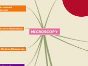
MICROSCOPY
Microscope is an instrument to observe microorgnisms
Subtopic
Light Microscope and Electron Microscope
Types of Staining
Differential Staining
Special Staining
Negative Staining
Endospore Staining
Flagella Staining
Microscope Parts
Base
Arm
Light Source
Subtopic
Coarse Adjustment Knob
Fine Adjustment Knob
Stage
Stage Clips
Substage
Nosepiece
Objectives
Body Tube
Permits greater resolution
UV Microscope
Has shorter wavelength
Confocal Micoscopy
Creates a sharp 3D image bybusing laser beam nd numerous applications including study of biofilms
Dark Field Microscope
Produce a bright image of the object
Used to observe internal structures in eukaryotic micoorganisms
Used to identify bacteria
Phase Contrast Microscope
Converts differences in refractive index
Excellent way to observe living things
Stain is not necessary
Fluorescence Microscope
Expose specimen to ultraviolet, violet or blue light
Specimens usually stained with flourochromes
Applications in medical microbiology
Electron Microscope
Employs a beam of electron in place pf light wave to produce magnified image
To study the surface features of cells and viruses
Preparations of Specimens for Light Microscopy
Wet Mount
Demonstrating motility of microorgansims
Smears
Preparing Smears
Fixation
Staining
UNITS OF MEASUREMENT
Metric System
Micoorgansims and their structural components are measured in micrometers, nanometers and angstroms
