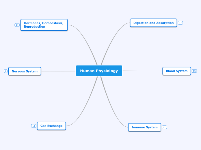
Human Physiology
Gas Exchange
Antagonistic Muscles
Muscles only do work when they
contract, shortening. By relaxing,
the muscles become longer. If
movement in two directions is
required, two different muscles
must work against each other.
Airways and Pressure
Pressure Changes
Muscle contractions cause the
pressure changes inside the thorax
that force air in and out of the lungs
to ventilate them.
Airways
Air is carried to the lungs in the
trachea and bronchi and then to
the alveoli in bronchioles.
Pneumocytes
https://youtu.be/RjWT0eNw40o
Type II
Creates the moist surface inside
alveoli to keep the sides from sticking
to each other. They create a mono-
layer with hydrophilic heads facing
water and hydrophobic tails facing air.
This reduces surface tension and
prevents lung collapse.
Type I
Thin alveolar cells adapted to carry out
gas exchange. Lunges have large surface
area alveoli covered in mostly Type I
pneumocytes. The distance from these to
the blood capillaries is very low, so the
gas exchange has little distance to diffuse
across.
Ventilation
https://youtu.be/IMDEXGM-87s
Maintains concentration gradients of
oxygen and carbon dioxide between
air in alveoli and blood flowing in
adjacent chambers. Gases exchange
because of a concentration gradient.
Nervous System
Synapses
https://youtu.be/W4N-7AlzK7s?t=204
Synapses are junctions between neurons
and receptor cells. Neurotransmitters send
signals across synapses. When pre-
synaptic neurons are depolarized, they emit
neurotransmitters into the synapse. This
causes Ca+ ions to diffuse through the
membrane into the neuron. This influx of
positive ions releases neurotransmitters via
exocytosis, which then bind onto receptors
on the post-synaptic neuron. When this
happens, the sodium ion channels open and
diffuse against their concentration gradient.
This triggers the action potential and sends
the signal down the neuron.
Neurons
https://youtu.be/W4N-7AlzK7s?t=92
Transmit electrical impulses from
dendrites to terminal buttons. There
is a resting potential caused by the
pumping sodium and potassium
across their membranes. This creates
an area of positive ions on the outside
and an area of less positive ions on
the inside. The resting potential lets
the neuron re-polarize or depolarize,
sending the signal down the neuron.
Any electrical signal of above -70 mV
sends it along the neuron.
Hormones, Homeostasis,
Reproduction
Menstrual Cycle
Chemical Effects
https://medium.com/@bicspuc/menstrual-cycle-an-important-process-of-human-reproduction-e22a4abce2e2
Luteal phase
https://youtu.be/QfjiOZ-iCeA?t=384
The wall of the follicle that released
the egg becomes a body called the
corpus luteum. If fertilization does
not happen, it breaks down. The
lining of the uterus is also broken
down and shed.
Follicular phase
https://youtu.be/QfjiOZ-iCeA?t=94
An egg is stimulated to grow in each
follicle at the same time the lining of
the uterus is repaired and begins to
thicken. The follicle most developed
releases the egg into the oviduct.
Controlled by negative and positive
feedback mechanisms.
Sex Determination
https://youtu.be/D2hVgujy2E8
Females
Estrogen and progesterone cause prenatal
development of female reproductive organs
and female secondary sexual characteristics
during puberty.
Males
Testosterone causes prenatal development
of male genitalia and both sperm production
and development of male secondary sexual
characteristics during puberty.
A gene on the Y chromosome
develops testes and testosterone.
Hormones
Melatonin: controls circadian rhythms.
Leptin: inhibits appetite.
Thyroxin: regulates metabolic rate
and body temperature.
Blood Glucose Control
https://youtu.be/WVrlHH14q3o?list=PLubGh95H6twxSvUL8LRtz0qsuj_LK5XCv&t=430
Alpha and beta cells in the pancreas
respond to changes in blood glucose
levels. When this happens, insulin and
glucagon are released. Insulin turns
glucose into glucagon, lowering the
level of glucose in the blood stream.
This is stored in the liver, and is
released when levels get too low. It is
then turned back into glucose, bringing
the body back into homeostasis.
Immune System
Immune Response
Specific
https://youtu.be/XeOmXQSQl2E
1. Invader enter body (pathogen)
2. Detected and trigger immune response (antigen)
3. Pathogens are ingested by macrophages.
4. Antigens are displayed on cell membranes.
5. Helper T-cells specific to antigen are activated by
macrophages.
6. B-cell specific to the antigen is activated by
proteins from helper T-cells.
7. B-cells divide repeatedly to produce antibody
secreting cells and memory cells
8. Antibodies attach to antigen and aid in its
destruction.
9. Memory T-cells remain to detect antigen in the
future.
Non-specific
In the event of a pathogen entering
the body, phagocytes form the next
level of defense. The pathogens then
absorb and break down the infections,
destroying them.
Blood Clotting
https://www.wfh.org/en/page.aspx?pid=635
1. Skin gets cut, blood vessels are
severed and begin to bleed.
2. Clotting begins if platelets release
clotting factors. If this does happen,
thrombin starts converting fibrinogen
into fibrin.
3. The fibrin forms a mesh net that
traps platelets and blood vessels,
creating a gel initially, that hardens
into a scab.
Primary Defense
Skin and mucus act as the
first line of defense against
infectious disease.
Blood System
The Cardiac Cycle
https://www.thoughtco.com/phases-of-the-cardiac-cycle-anatomy-373240
An electrical impulse travels from the S.A. node
to the A.V. node. The atria depolarizes, causing
them to contract. The signal then travels down
Bundle of His and separates into the left and
right bundles. The L. posterior and anterior go
to the front and back, respectively. The signal
uses Perkinje fibers to depolarize the ventricles,
while the atria simultaneously re-polarizes.
Heart Structure
https://www.news-medical.net/health/Structure-and-Function-of-the-Heart.aspx
Blood Vessels
https://youtu.be/v43ej5lCeBo
Capillaries
Permeable walls allow the blood to
exchange materials between the
cells in the tissue and the blood in
the capillaries. The walls are made
up of one layer of thin endothelium
cells with pores to be permeable.
Veins
Collect blood from tissues at a low
pressure and bring it back to the
atria. They are thin walled because
the blood is at a much lower pressure
and use gravity and skeletal muscles
to create blood flow. Valves prevent
any backflow from occurring.
Arteries
Convey blood from the heart to the
tissues of the body. They are thick,
strong and are able to withstand
high pressures.
Digestion and Absorption
Absorption
https://byjus.com/biology/absorption-of-digested-foods/
Different methods of absorption:
Passive Transport
Facilitated Transport
Active Transport
Simple Diffusion
Villi and Micro Villi
https://youtu.be/2YuEz8P98VM
The villi and microvilli exist to
provide more surface area for
digestion. This allows more
absorption to take place, as
well as creating more places for
molecules to be broken down
by enzymes.
Digestion in the Small Intestine
The walls of the small intestine
have many enzymes that further
break down the food. Because
the molecules must be broken
down to a tiny size before being
absorbed into the villi, digestion
in the small intestine takes hours.
Enzymes digest the majority of
macromolecules. For example,
protein chains are broken down
into smaller chains by protease.
The pancreas releases enzymes
into the lumen of the small
intestines.
Peristalsis
https://www.youtube.com/watch?v=kVjeNZA5pi4
The contractions of the small
intestines prevent food from going
back to the mouth and are
continuous in one direction.

