RNA
RNA is single stranded and is composed of a phosphate, ribose sugar, and nitrogenous base. RNA also forms phosphodiester bonds to link RNA monomers together in a 5' to 3' direction.
Genetic Information
DNA
A DNA monomer, also known as a nucleotide is comprised of a Phosphate, deoxyribose sugar, and nitrogenous base. The phosphate forms a phosphodiester bond with the nucleoside(sugar-base) on the 3' and 5' ends of the 5 carbon sugar extending in that direction. The DNA molecule is composed of an anti-parallel double helix strand that connects the base pairs(A-T,C-G) via Hydrogen bonds.
The tRNA's job is to carry the correct amino acid, which is then chained together.
A sequence of about 20
amino acids at or
near the leading end
(N-terminus) of the
polypeptide
New protein entering
endomembrane system
Protein
Protein synthized
(translation)
started in cytoplasm
signal peptide
marked protein
signal-recognition particle(SRP)
A protein-RNA complex that
recognizes signal peptide as
emerges from ribosome and
helps direct ribosome to ER
by binding to a receptor
protein on the ER.
receptor protein on the ER membrane
Signal peptide
SRP
part of the mutliprotein
translocation complex
that forms a pore.
polypeptide
targeted to function as
membrane protein
Both travel to the
plasma membranve in
a trasport vesicle
Destinations of proteins
synthesised on free ribosomes
secreted outside the cell
ER lumen
Final Processed MRNA is then exported into Cytoplasm
This is made up of a single RNA strand about 80 nucleotides long. The strand base pairs with itself in three different sections forming a clover like shape when flattened into one plane (it's L shaped when fully folded), The bottom section of the tRNA contains the anticodon (3' to 5'), which binds to the codon (5' to 3') in the mRNA. The 3' end is the amino attachment site. The anticodon is antiparallel to the mRNA.
similarities in both
Require Transcription Factors
Require Promotor Region
Translation
These have two subunits composed of RNA and proteins.
60S and 40S (80S total)
50S and 30S subunits (70S total)
An amino acid and its matching tRNA enter the active site of the synthetase enzyme determined by the amino acid.
The synthetase catalyzes the binding of the amino acid to its complementary tRNA through a covalent bond. AMP and two inorganic phosphate groups.
The tRNA- amino acid pair is released by the synthetase.
After mRNA leaves the nucleus, the smal ribosomal subunit is attached to the 5' cap to it. This occurs when the subunit recognizes a specific nucleotide sequence on the mRNA.
A bit further uptream of the start codon, an initiator tRNA, which contains the anticodon UAC, base pairs with the start codon (AUG). Note this tRNA carries methionine.
The large ribosomal subunit arrives, which completes the initiation complex. Initiation factors are used to bring all the components together and the hydrolysis of GTP provides the neccessary energy for assembly. Note that this first tRNA-amino acid complex goes into the P site of the large ribosomal subunit.
Codon Recognition
A new aminoacyl tRNA containing the complementary mRNA codon will base pair with the mRNA and go into the A site. Note that there are many aminoacyl tRNA's present but only the one with the right anticodon will bind to the mRNA.
An rRNA molecule of the large ribosomal subunit catalizes the formation of a peptide bond between the two amino groups in the A and P sites. Both amino acids will go on the A site tRNA and form a chain of amino acids.
Peptide Bond Formation
The now empty tRNA in the P site will then move over to the E site and the tRNA and amino acid chain will move over to the P site. This is called translocation.
Translocation
Termination
A release factor will bind to the stop codon in the mRNA and will go into the A site. This will then use GTP to release all the components of the translation complex.
Transfer RNA
Cell Transcription
Eukaryote replication
Forms PRE-MRNA
G-cap
Poly-a Tail
Poly-A Polymerase
Exons
Introns
Spliceisome
Uses RNA Polymerase 2
Prokaryotes replication
Forms MRNA
Series of RNA Nucleotides, ready to be translated
Uses RNA Polymerase
Cellular Response
Once activated, relay proteins are able to bring about responses in the cell.
Relay Proteins
After autophosphorylation, inactive relay proteins are able to bind to the phosphate groups attached to each tyrosine. These relay proteins then become activated by taking the phosphate group and detaching from the tyrosine. Once the relay proteins take away the phosphate group from tyrosine, the tyrosine becomes deactivated.
Autophosphorylation
After dimerization, the tyrosines on one side of the kinase take a phosphate group from ATP, forming ADP, and add it to a tyrosine on the other side of the kinase. Said other side, does the same. Once every tyrosine is bound to a phosphate group the kinase is considered fully activated.
Second Messengers
Small, non-protein. water-soluble molecule or ion, suck as a Cyclic AMP(cAMP), that relays a signal to a cell's interior response to a signaling molecule bound by a signal receptor protein. Messengers can spread thoughout the cell by diffucion.
Transduction is a step or series of steps that converts the signal to a from that can bring about a specific cellular response. Transduction usually involves a signal transduction pathway, sequence of changes in a series of different molecules.
Phosphorylation Cascade
Series of chemical reactions during cell
signaling mediated by kinases, the kinase
turn phosphylates and activates another,
leading to phosphorylation of other proteins.
Active Protein Kinase 1
Active Protein Kinase 2
Inactive Protein Kinase
Active Protein
Protein Phosphatase(PP)
Inactive Protein
Cellular response
Intercellular receptors
Formation Of MRNA
Forms specific proteins
Cell response
Extracellular receptors
Tyrosine Kinase Receptor
Receptor Tyrosine Kinase Proteins
Dimerization
When a signal molecule binds to each of the inactive monomers' binding site, the two tyrosine kinase proteins dimerize, this means that the mirrored monomers join together and form a tyrosine kinase dimer.
Tyrosines
There are 3 of these attached to each protein, these become activated and deactivated and are able to interact with relay proteins.
Alpha Helix
This section of the receptor is embedded within the membrane itself, keeping the active site on the outside of the cell and the tyrosines on the inside of the cell.
Signal-Binding Site
Signal molecule binds to the signal binding site. This receptor is found outside of the cell membrane. This changes shape when it's activated/deactivated.
GPCR
G protein
GDP and GTP
GDP
GTP
Adeynl Cyclase
CAMP
Protein Kinase 1
ATP
ATP Synthesis
Forms ATP!!
Photophosphorylation
Similar to Oxidative phosphorylation in cellular respiration, Photophosphorylation is powered by a H+ ion concentration gradient in which H+ ions are pumped from the stroma into the thylakoid space where there is a high concentration gradient of H+ ions. The H+ protons are then pumped back out to the stroma through the ATP synthase protein where ATP is generated via chemiosmosis.
Substrate-level Phosphorylation
Substrate-level Phosphorylation occurs in glycolysis and citric acid cycle, where the given substrate provides a phosphate to ADP which is attached to the enzyme, resulting in the formation of ATP and a new product. Examples of given substrates are 1-3 Biphosphoglycerate and PEP in glycolysis; Succinyl CoA in the citric acid cycle.
Oxidative Phosphorylation
In oxidative phosphorylation, the electron carriers NADH and FADH2 become oxidized as they lose their electron, ultimately transferring to Oxygen where water is formed. It's respective proton is pumped via active transport through the protein H+ pumps where it creates a high concentration gradient of H+ ions in the intermembrane space. The H+ ions are then pumped back into the matrix via chemiosmosis as it moves down its concentration gradient powering the ATP synthase protein where ADP and P react to form ATP.
ADP
Cell Signaling
Cell signal stages
Reception
Transduction
Response
Local Signaling
Synaptic Signaling
Paracrine Signaling
Long Distance signaling
Group 6 Mind Map
by Joanna, Maximo, Silverio, Christian
The R-groups associated with Transmembrane proteins are orientated so that the n-terminus(partially positive) is outside the cell membrane and the carboxyl c-terminus group(partially negative) is inside the cytoplasm.
These types of proteins are inserted into the membrane. They can be partially or fully inserted.
Common organelles of Prokaryotes
Gas vacuole
Provides buoyancy for floating in aquatic environments
Endospores
Bacteria only feature. These form when there are unfavorable environmental conditions. Forms a copy of its chromosome, and surrounds itself with a multilayer cell wall
Pili
Long projections that form a channel between cells. Can transfer DNA from one cell to another
Flagellum
Helps cells be more motile, can be scattered or concentrated
Slime Layer
A sticky layer on the outside of the cell wall that is diffuse and adheres to substrate or other individuals
Plasmid
DNA that is separate from the main bacterial chromosome. BACTERIA Only
Fimbraie
Short Hair like projections, used to stick to substrates or other cells.
Capsule
A sticky layer on the outside of the cell wall that is dense and adheres to substrate or other individuals
Cell Wall
Wall outside the membrane, provides support ,protection and maintains cell shape. In bacteria this wall is made of Peptidoglycan
Complexes that synthesize proteins
Nucleoid
Area where the chromosome( DNA) is located in prokaryotes, is not surrounded by a membrane
Animal cell organelles/ Structures
Centrosomes
Centrosomes are the factory for Microtubules. Within a centrosome there are two centrioles which help in chromosome movement in cell divison.
Extracellular Matrix
The animal version of a cell wall. the ECM is composed of various different proteins. Such as fibronectin,peptidoglicyans and collagen. Components of the ECM bind to proteins called Integrins( in the cell membrane) and they are attacked to microfilaments. Changes in the ECM can trigger changes inside the cell
Higher proportion of
unsaturated
Viscous
The membrane would be more viscous
as the packed hydrocarbon tails would
increase the membranes viscosity. And
because the tails are pack together
they are saturated.
A phosphate group will attach to an OH in glycerol in order to create the phospholipid
Organelles of plant cells
Cell wall
Outer layer, that maintains cell shape, and protects from mechanical damage. Made from polysaccharides and proteins, and cellulose
Central Vacuole
Prominent organelle in older plant cells, functions include storage, breakdown of waste, hydrolysis. Enlargement of the vacuole is a major part of plant growth
Chloroplasts
A Plastid that contains a inner and outer membrane, is composed of sacs called Thylakoids, Groups of thylakoids are called granums. Converts solar energy to Chemical energy
Fluid
The membrane would be more fluidity
as the hydrocarbon tails would not
allow the membrane to have a stable
form. As the tails are kinked
they are unsaturated.
Amphipathic molecule
This molecular orientation maximizes contact of the hydrophilic regions of a protein with water in the cytosol and extracellular fluid, while providing its hydrophobic parts with a nonaqueous environment.
Hydrophobic region
The tails are in contact
other tails and not water.
No affinity for water
"fears water"
Hydrophilic region
The hydrophillic head
are in contact with water and
other aqueous solutions.
Affinity for water
"loves water"
Move sideways within membrane
Phospholipids Bilayer
Held together primarily by hydrophobic interactions
Hydrophobic Tail
Hydrphilic Head
Specific phase transition temprature
Below this temperature
Rigid
gel phase
Above this temperature
Liquid
Liquid crystalline phase
Type of hydrogarbon tails
Saturated Hydrocarbon tails
Tails are packed together
Unsaturated hydrocarbon tails
Tails are kinked
The tails can not pack together
and are loose.
Modes of Transport
Vesicle Transport
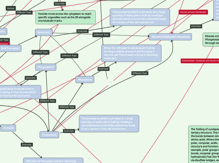
Vesicles move across the cytoplasm to reach specific organelles such as the ER alongside microtubulin tracks.
Ion Channels
Membrane proteins that allow for transport of ions across membranes
Gated Channels
Voltage-Gated
Open and close in response to changes in membrane potential.
Ligand-Gated
Open and close when a neurotransmitter binds to a channel.
Stretch-Gated
Open when membrane is mechanically deformed, they sense stretch.
These ion channels open and close in response to stimuli.
Ungated
These ion channels are always open
Bulk Transport
Exocytosis
The process by which a cell takes out a large quantity of molecules in bulk by creating a vacuole/vesicle around the substances made of a section of the cell memebrane.
Endocytosis
The process by which a cell takes in a large quantity of molecules in bulk by creating a vacuole/vesicle around the substances made from a section of the cell membrane.
Receptor Mediated Endocytosis
A specialized type of pinocytosis which allows a cell to bring in specific substances by creating coated vesicles around specific molecules when the proper ligand binds to a receptor found on the outside of the cell membrane, which alerts the cell when the necessary substance is present.
Pinocytosis
When the cell takes in extracellular fluid by forming a vesicle around it and bringing it into the cell. This is used to bring in dissolved molecules.
Phagocytosis
When a cell engulfs large food particles or other cells by creating a food vacuole which is digested after fusing with lysosomes.
Active Transport
Electrogenic Pump
Proton Pump
H+/Sucrose Constransporter
Energy generated by the proton pump drives the active transport of nutrients in the cell. In this case, sucrose is moved against the concentration gradient using energy supplied by the proton pump.
H+ is transported across the concentration gradient, ATP is used to facilitate this. As the positive charge leaves the cell, a negative charge develops inside the cell and protons want to come back in the cell.
A transport protein that generates a voltage across a membrane, otherwise known as membrane potential.
Allows substances to move from areas of low to high concentration in order to maintain a high concentration. This type of transport requires energy
Passive Transport
Osmosis
Diffusion of free water across a selectively permeable membrane. It moves from an area of low SOLUTE concentration to high SOLUTE concentration until the SOLUTE concentration is even on both sides of the membrane.
Diffusion of a substance across a membrane without the use of energy
Facilitated Diffusion
Passive transport aided by proteins.
Simple Diffusion
The movement of molecules across the membrane without the help of energy or proteins. They move from areas of high concentration to areas of low concentration.
Cotransport
This occurs when active transport of a solute indirectly drives transport of other substances.
Water Balance of Cells
Hypotonic
When the solute concentration is lower outside the cell than inside the cell, the cell gains water and expands. This is the normal state for a plant cell (turgid). This is known as a lysed state in animal cells.
Hypertonic
When the solute concentration is greater outside the cell than inside the cell, the cell looses water and shrivels up. This is a shriveled state in animal cells and a plasmolyzed state in plant cells.
Isotonic
When the solute concentration is the same outside and inside the cell, there is no net movement of water. This is the normal state for animal cells. In plant cells this is known as a flaccid state.
Tonicity
The ability of a surrounding solution to cause a cell to gain or lose water.
Channel Protein
This transmembrane protein allows water molecules or other small molecules to pass through the membrane via a channel.
Carrier Protein
Sodium Potassium Pump
3 Na+ ions found in the cytoplasm binds to the pump when it's open facing the inside of the cell. This stimulates phosphorylation by a kinase using ATP, which leads to the protein changing shape and opening towards the outside of the cell, releasing the 3 Na+ ions and allowing 2 K+ ions to bind. After phosphorylation, the loss of the phosphate group allows causes the protein to return to its original shape, which has a lower affinity for K+, releasing the 2 K+ ions.
This transmembrane protein transports substances from one side of the membrane to another by changing shapes.
Phospholipid
Polarity
Phospholipids are amphipathic, meaning they have a hydrophobic and hydrophilic side. The hydrophobic side is the tail end, which contains the lipids. The carbon chains attract each other though hydrophobic and Van der Waals interactions. The hydrophilic side is the head, which contains the positively charged phosphate group. The charge in this group allows the head to interact with water through hydrogen bonds. When placed in water, the hydrophobic tails face each other, forming a bilayer made up of two rows of phospholipids, with the tails facing inwards, and the heads facing outwards.
Phosphate Linked Head
Fatty Acids
These have a carboxylic acid on one end and are made up of a carbon chain surrounded by hydrogens everywhere except where there is a double covalent bond between carbons. The types of fatty acids presents determine the permeability of the membrane, with the presence of unsaturated fatty acids increasing permeability and vice versa.
Unsaturated
These occur when there are double covalent bonds between carbons present in a fatty acid. They come from plant sources and are liquid at room temperature. These also don't have hydrogen atoms along the carbon chain where double bonds are present.
Saturated
These occur when there are no double covalent bonds between carbons in a fatty acid. They are commonly found in animal sources and are solid at room temperature.
Glycerol
This is linked to two fatty acids in a phospholipid. It's linked through an ester bond which is formed through a dehydration-condensation reaction. The third OH in glycerol attaches to the phosphate head.
Primary Structure
Peptide bonds hold together monomers of amino acids in primary structure. The peptide bond occurs between the partial positive amino group and the partial negative carboxyl group via dehydration synthesis reactions.
Protein Structures
Quaternary Structure
Quaternary Structure is formed between two tertiary structures. A common quaternary structure is hemoglobin.
Tertiary Structure
The folding of a polypeptide forms a 3D tertiary structure. This folding is caused by the bonds between similar side chains in amino acids. Where the types of R-groups: polar, nonpolar, acidic, basic affect the structure and function of the protein. For example, polar groups bond via hydrogen bonds, nonpolar groups bond via hydrophobic/Van Der Waals interactions or via disulfide bridges, and basic/acidic R-groups form ionic bonds.
Secondary Structure
The bond that occurs in secondary structure are Hydrogen bonds between the lone hydrogens in the amino acid monomer. Secondary structure orientates the amino acids into beta-pleated sheets or alpha helix structures.
Membranes
Organelle Membranes
Organelle made of different sacs, each with its own membrane, these can fuse together.
Peroxisomes
These have a single membrane.
Chloroplast
This contains an inner and outer membrane as well as other membranous sacs called thylakoids.
This has a double membrane made up of an outer membrane and an inner membrane which is folded into cristae. In between the two is a space called the intermembrane space.
Their membranes help keep the acid hydrolases within the organelle. They contain a protein pump in their membrane because the more protons it has the more acidic the environment becomes. Its cell membrane can also fuse with another lysosome if it's defective, where it is then broken down. This process is called autophagy
Endoplasmic Reticulum
Rough ER has ribosomes around its membrane and smooth ER doesn't.
Double membrane that has nuclear pores embedded in in, which are lined with proteins called porin proteins that help transport molecules in and out of the nucleus.
Phospholipid Bilayer
This is a gel-like substance in cells that suspends organelles and is a medium for chemical reactions.
Extracellular Fluid
ECM
This is the environment that surrounds the cell. This is usually aqueous and therefore polar, causing the hydrophobic tails of the phospholipids to face away from it.
Membrane Proteins
Attachment to Cytoskeleton and ECM
Microfilaments, or any part of the cytoskeleton, can be bound to membrane proteins noncovalently. This helps maintain the cell's shape and stabilizes the location of different proteins within the membrane. Likewise, proteins can also bind to ECM molecules in order to make extra and intracellular changes.
Intercellular Joining
Membrane proteins of adjacent cells can join together through gap, tight, and other types of junctions, much like in cell-cell recognition, but the bond lasts longer.
Cell-Cell Recognition
Some glycoproteins can serve as identification tags which are recognized by the membranes of other cells, allowing cells to recognize, communicate with, and count, cells of the same type, or in some cases, different types of cells.
Signal Transduction
This occurs when a membrane protein called a receptor has an active, or binding, site with a specific shape that's unique to that protein. This shape allows for a signaling molecule with a matching shape to fit in place and convey a message to the cell by binding to the receptor, much like a key going into a lock.
Enzymes
Kinases activate inactive enzymes and Phosphatases inactivate active enzymes through transfer of phosphate group.
Enzymes, a type of protein that speeds up chemical reactions, may be present along the membrane. These have an active site to which a reactant binds. There may be several enzymes that form a chain reaction in order to carry out steps of a metabolic pathway.
Transport
Channel proteins are used to move substances from one side of the membrane to another through facilitated diffusion or active transport.
Integral Proteins
Transmembrane Proteins
Peripheral Proteins
These types of proteins are anchored to the membrane.
Cholesterol
A type of steroid present in animals. It's a common component of membranes which regulates permeability. For instance, if a membrane has too many unsaturated fatty acids and is too permeable, cholesterol would help reduce the permeability of the membrane. On the other hand, if too many saturated fatty acids are present, cholesterol could help increase permeability.
Mitochondria
Powerhouse of the cell, organelle which conducts cellular respiration, and where most atp is generated. Contains double membrane. As well as DNA and ribosomes in its matrix
Rough Endoplasmic reticulum
Rough Er, is studded with ribosomes( Bound ribosomes) Secretes glycoproteins, transport vesicles and is the membrane factory
Smooth Endomplasmic Reticulum
Synthesizes lipids, metabolizes carbs, and detoxifies drugs and potions.. also stores K+
Free Ribosomes
Free Ribosomes are suspended in the ribosome, they function within the cytosol, meaning they do work mostly inside the cell. They are the first enzymes that catalyze sugar breakdown
Bound Ribosomes
bound ribosomes can be found in the E.R. These tend to create proteins that are either secreted out of the cell or membrane proteins. Both bound and free are identical and can alternate jobs
Microfilaments
Intertwined actin strands, Primary functions are; that maintain cell shape, changes in cell shape, muscle contraction, cell motility, and cell divison. is able to cause contractions through Myosin protein.
Intermediate Filaments
Fibrous proteins that are supercoiled into thicker cables. Composed of different types of proteins from the Keratin family. Main functions involve maintaince of the cell shape, anchorage of nucleus and other orgnalles, and the forms the nucleus lamina. These filaments are in between microtubules and Microfilaments in size, hence the name. NOTE: THESE ARE NOT IN PLANT CELLS, BUT I THOUGHT IT WAS INPORTANT TO HAVE ALL THREE TOGETHER IN ORDER TO VISUALIZE THEIR DIFFERENCES
Chromatin
Material consisting of DNA and protein, visible in dividing cells, as condensed chromosomes
Nucleolus
Nonmembranous structure involved in production of ribosomes, can be one more in a nucleus.
Common organelles in Eukaryotes
Lysosomes
Membrane bound organelle that is filled with enzymes. these enzymes promote hydrolysis of molecules. Will break down food , or foreign cells when cells commit phagocytosis. Will break down these particles into amino acids and simple sugars which are diffused into the cytosol. Can also break down defective organelles for recycling, this is called autophagy
Endoplasmic reticulum
Network of membranous sacs and tubes active in membrane synthesis and other metabolic processes. has two parts a rough ER and smooth ER
Perioxisome
organelles that is in a vesicle that is full of enzymes. These enzymes extract hydrogen atoms from molecules( Usually bad) and convert it to H2O2, and then into H2O
Ribosomes
Cytoskeleton
Microtubules
Hollow tubes that are made up of Alpha Tubulin and beta Tubulin. Its main functions are maintacie of cell shape, cell motility, chromosome movements during cell division and organelle movements. Kind of like a railroad in the cells
Golgi Apparatus
Organlles that is the distributing center of the cell. Active in synthesis, modification, sorting, and secretion of cell products outside the cell.
Nucleus
Nuclear Envelope
nuclear pores
Small pores that Allow transport into and out of the nucleus.
A double Membrane, that encloses the nucleus. Continous with the endoplasmic reticulum
Common Structures in both Prokaryotes and Eukaryotes
Cell Membrane
Selectively Permeable barrier, separates interior of cell from the exterior.
Cytoplasm
Cytoplasm is composed of cytosol, a gel like substance that surrounds the organelles in the cells.
Prokaryotic cells
The domains of life
Bacteria
Archaea
Eukarya
Eukaryotic Cells
Animal cells
Plant Cells
O
Main topic
