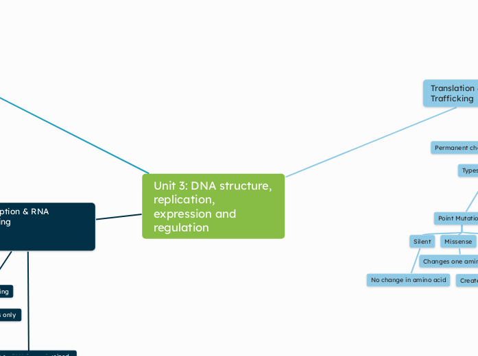
Unit 3: DNA structure, replication,
expression and regulation
Transcription & RNA Processing
RNA processing
Eukaryotes only
5' Cap
Happens as RNA is synthesized
Protects mRNA
3' Poly-A tail
Cleavage factor binds
Cut RNA downstream
Poly-A Polymerase adds A LOT of A's to 3' end
~100-300
Splicing
Removes introns
Joins exons
Splicesome
snRNA + proteins
Alternative splicing
makes many proteins from one gene
Transcription
Prokaryotes
Location
Cytoplasm
Transcription and Translation coupled here
Initiaton
promotor = specific sequece on DNA
RNA polymerase binds to promotor
Unwinds DNA
Forms transcription bubble
Helicase unwinds DNA at promotor
Topoisomerase keeps it from rewinding and relieves stress
Elongation
RNA polymerase adds complementary nucleotides
A-U
C-G
RNA synthesis in the 5' --> 3' direction
Termination
Termination factor reached
Newly synthesized mRNA released
Eukaryotes
Location
Nucleus
goes to cytoplasm for translation
Initiation
Promotor region
Promotor = TATA box
Enhancer sequences
Transcription factors bind to promotor sequnce
recruits RNA Polymerase II to bind to promotor
Helicase unwinds DNA at promotor
forms transcription bubble
Topoisomerase keeps it from rewinding and relieves stress
Elongation
RNA polymerase reads 5' --> 3'
synthesizes complementary RNA 3' --> 5'
U --> T in RNA
Termination
RNA polymerase reads termination factor
Cleavage factors cut RNA transcript
RNA polymerase detaches
DNA Structure & Replication
Double helix model (Watson & Crick)
Antiparallel strands (5′→3′ / 3′→5′)
Sugar-phosphate backbone
Nitrogenous bases: purines (A, G) vs pyrimidines (T, C)
Complementary base pairing: A–T (2 H-bonds), G–C (3 H-bonds)
Phosphodiester bonds (covalent, between nucleotides)
Hydrogen bonds (between bases)
Enzymes in DNA Replication
Helicase – unwinds DNA
Topoisomerase – relieves tension ahead of fork
Single-Stranded Binding Proteins (SSBPs) – stabilize separated strands
Primase – adds RNA primers
DNA Polymerase III – synthesizes new strand (5′→3′)
Sliding clamp – increases DNA pol processivity
DNA Polymerase I – removes RNA primers & fills gaps
Ligase – seals nicks between Okazaki fragments
Overview
Semiconservative replication model
Meselson-Stahl experiment (14N vs. 15N labeling)
Replication origin (ORI)
Replication fork
Bidirectional replication
Replication bubble
Leading vs Lagging Strand
Leading strand synthesized continuously
Lagging strand synthesized discontinuously
Okazaki fragments
Directionality matters: DNA pol adds only to 3′ end
Features of Replication
High speed (E. coli: 2000 nt/sec)
High fidelity (1 error per 10⁶ bases, proofreading)
Requires dNTPs as substrates
Polymerases need a primer to begin synthesis
Griffith's transformation experiment
Hershey & Chase bacteriophage experiment
DNA vs. protein as genetic material
Chargaff's rule (A = T, C = G)
Translation & Protein Trafficking
mRNA -> Protein
Eukaryotes
mRNA Processing: Capped, spliced, poly-A-Tail
larger ribosomes
Prokaryotes
No mRNA processing
smaller ribosomes
Steps of Translation
1. Initiation
Small ribosomal subunits bind to mRNA at start codon (AUG)
tRNA carries methionine to start codon
Large ribosomal subunit joins to form complete ribosome.
2. Elongation
Ribosome moves along mRNA, reading codons.
Matching tRNAs bring amino acids.
Amino acids are joined by peptide bonds
3. Termination
Ribosome reaches a stop codon (UAA, UAG, UGA).
Release factor binds
Ribosomal subunits separate.
Secretory Pathway
Signal peptide directs ribosome to ER membrane.
Protein enters ER lumen ).
Travels to Golgi Apparatus in vesicles.
Golgi further modifies, sorts, packages.
Final vesicle sends protein to:
Plasma Membrane
Lysosome
Cel Membrane
Permanent change in DNA sequence
Types of Mutations
Point Mutations
Silent
No change in amino acid
Missense
Changes one amino acid
Nonsense
Creates premature stop codon
Frameshift Mutations
Insertion
Deletion
Reading frame shift
Two types of Ribosomes
Free Ribsomes
Bound Ribosomes
