venous filter
Patients with high risk of pathology recurrence
Floating emboli in the inferior vena cava
Patients contraindicated for anticoagulation
transesophageal echocardiogram
90%
Pulmonary hypertension
systolic dysfunction of the right ventricle
Paradoxical movement of the interventricular septum
60%
Clots are carried through the bloodstream
Hypercoagulability
Stasis
Describes a triad
Rudolph Virchow
Endothelial damage
The presence of clots in the veins of patients suffering from sudden death
Giovanm Basttista
A process of blood clotting function without identifying the mechanism
French surgeon
ayurveda medicine
600-100 B.C.
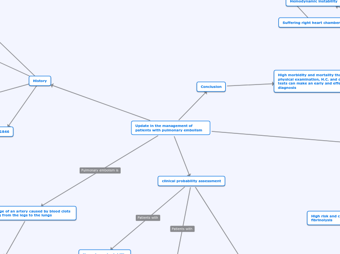
1846
1576
1761
Update in the management of patients with pulmonary embolism
Conclusion
High morbidity and mortality than with physical examination, H.C. and diagnostic tests can make an early and effective diagnosis
Treatment
Pregnant patients
Suffering right heart chambers
Hemodynamic instability
Diagnostic means
TAC scan of the chest on pulmonary angiography
Greenfield filter infrarenal position
Surgical pulmonary embolectomy
persistent hemodynamic instability
High risk and contraindication for fibrinolysis
After the procedure is performed
Normotechnical cardiopulmonary bypass
Mass pulmonary embolism
Streptokinasa 250.000 UI in
the first 30 minuts
100.000 UI every hour for 24 hours
Anticoagulation
Alteplasa 100 mg in the first 2 hours
Scheme that decays due to the change to the accelerated administration mode
1,5 millions of units in 2 hours
Pulmonary embolism
It is managed outpatient when
Low risk
Treated with
HBPM
External consultation
hemodynamic stability
clinical probability assessment
Criteria to use
Ginebra
Low
0-3
Take Dimero D's lab
Biological marker product of fibrin degradation
It is not reliable for the diagnosis of embolism, it is part of the diagnosis of
Peripheral arthropathies
Sepsis
Nephropathies
Trauma
8 Hours
Intermediate
4-10
High
Greater than 10
Wells
Evaluate
clinical judgment
Signs and symptoms
Clinical probability
Predisposing factors
Unlikely
Greater than 4
Probable
De 0 a 4
hemodynamically stable
Tomography
Hemodynamic stability
Dimero D
Multislice tomography
History
Blockage of an artery caused by blood clots moving from the legs to the lungs
Clinical presentation
variability from
Shock
Syncope
Haemoptysis
Tachypnea
dyspnoea


