Gene Expression
Regulation
regulation @ transcription initiation
enhancers: sequences on DNA in which are far away from the gene
uses transcription factors to bind onto promoters: activators & repressors
repressors: decreases level of transcription = less mRNA
activators: increases level of transcription = more mRNA
when activators are bound, it will cause the DNA to bend to bind to the mediator proteins to increase transcription
when different proteins are needed in different organs, one organ (ex. liver) may have an activator protein for one protein (ex. albumin) that the other organ (ex. eye) may not have; helps different organs have different functions from each other
DNA Replication
Steps of DNA Replication
7.) DNA Ligase links the Okazaki fragments together to create a single, new strand
6.) DNA polymerase I removes RNA primers and replaces them with DNA
5.) DNA polymerase III adds nucleotides to the 3' end of primers and continues elongation on both strands
4.) Primase adds the RNA primer to the template DNA so that polymerase can start replicating
Needs to continuously add primers to the lagging strand to make several Okazaki fragments
3.) Single Stranded Binding Protein (SSB) binds to the single stranded DNA and makes sure it stays apart
2.) Helicase binds and unwinds the two strands of the template DNA by breaking the hydrogen bonds
1.) Topoisomerase binds to the ORI and relieves strain due to DNA supercoiling
Leading & Lagging Strands
Lagging strand: discontinuous replication in the opposite direction of the replication fork movement
Okazaki fragments are eventually covalently combined by DNA Ligase
Replicates in multiple, small segments called Okazaki Fragments that each need and RNA primer
Leading strand: continuous replication in the same direction
of the replication fork movement
Only 1 RNA primer is required for the
replication of the leading strand
ORI & Replication Forks
Proteins bind to the ORI and separate the two strands of DNA,
forming a replication fork (or bubble)
DNA replication proceeds bi-directionally
Replication forks: Y-Shaped regions at
each end of the replication bubble where
DNA is unwound
DNA replication begins at specific DNA sequences called the
Origin of Replication (ORI)
Eukaryotes have multiple ORIs
Prokaryotes have 1 ORI
Components of DNA Replication
DNA Ligase
Covalently joins together Okazaki Fragments in lagging strands
DNA Polymerase I
Replaces RNA Primers with DNA
DNA Polymerase III
Builds new DNA strand using the old DNA strand as a template
Primase
Creates RNA Primers
Single Stranded Binding Protein (SSB)
Binds to and stabilizes single
stranded DNA
Helicase
Unwinds DNA double helix at the replication fork
Topoisomerase
Relieves DNA supercoiling ahead of replication fork
Transport
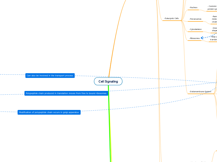
5.) after the Golgi, the protein will go to its target organelle or leave the plasma membrane
target organelles: ER, plasma membrane, lysosomes
4.) after the rough ER, the polypeptide will be received by the cis side of the Golgi apparatus, modifications take place throughout the Golgi, then will be transported from the trans side of the Golgi
3.) polypeptides produced in free ribosomes go to bound ribosomes on the rough ER
2.) mRNA goes to free ribosomes in the cytoplasm to start translation
polypeptides have destinations from free ribosomes: mitochondria, nucleus, peroxisomes, chloroplast, cytoplasm
1.) transcription happens in the nucleus to produce mRNA; mRNA leaves via nuclear pores
DNA Structure
Chargaff's Rule
Erwin Chargaff made two important discoveries about DNA:
1.) DNA base composition varies between different species
2.) For each species, the percent of A&T bases are roughly equal, as were the percents of the G&C bases
Meselson-Stahl Experiment
The parental strands separate and act as templates to synthesize new DNA that is complementary to it
Semi-Conservative Model: Replicated DNA molecules have one old/parental strand and one newly built strand
In 1958, Meselson and Stahl demonstrated that E-coli replicates DNA via the Semi-Conservative model
The Hershey-Chase Experiment
Used a bacteriophage to show that DNA was injected into the bacteria and proteins were left outside of the coat
In 1952, scientists Hershey and Chase used bacteriophages to confirm that DNA is the genetic material, NOT proteins
The Griffith Experiment
Overall conclusion showed that some unknown factor was
altering the bacteria. At the time, scientists did not know if this factor was DNA or proteins.
Showed that bacteria have the ability to transform genetic
material (S strain, R Strain, and Heat-Killed S Strain) and injected it into mice to test
Live R Strain + Heat-Killed S Strain
Heat-Killed S Strain
Live R Strain
The mouse lived
Live S strain
The mouse died
In 1928, Fredrick Griffith's experiment identified that some unknown genetic factor controls the traits of organisms
Overall Structure
DNA consists of two strands (double helix) of nucleotide monomers repetitively linked together
Free hydroxyl group at the 3' ends
Free phosphate group at the 5' ends
Transcription
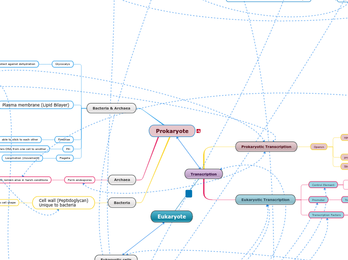
Eukaryotes (Nucleus)
Modification
A mature mRNA strand is formed.
The last process is called RNA splicing. A complex made out of RNA and proteins called spliceosomes splices certain sequences in DNA. Intron sequences are removed and exon sequences are joined together.
An enzyme called polyA polymerase adds a polyA tail (AAAAAA) to the 3' end of the pre-mRNA strand.
A guanine nucleotide modifies the 5' end of the pre-mRNA strand by adding a 5 CAP.
A sequence in the RNA called the polyadenylation signal sequence (AAUAAA) stops transcription. Proteins make a cleavage in pre-mRNA to release the strand.
RNA polymerase II reads the template strand (3' to 5') to make the RNA in the 5' to 3' direction. It adds nucleotides to the template strand to make RNA. It creates downstream.
RNA Polymerase II binds to the promoter with additional transcription factors to create the transcription initiation complex. It begins unwinding DNA at the transcription start point (+1) to begin RNA synthesis.
Proteins called transcription factors bind to the promoter before RNA Polymerase II.
Prokaryotes (Cytoplasm)
A sequence in RNA is the signal for the transcription process to stop. Once done, a mRNA strand is made.
RNA polymerase reads the template strand (3' to 5') to make the RNA in the 5' to 3' direction. It adds nucleotides to the template strand to make RNA. It creates downstream.
RNA polymerase unwinds DNA strand and start creating the RNA strand at the transcription start point. (+1)
RNA Polymerase binds to the promoter which is upstream.
Translation
Translation begins on free ribosomes (composed of rRNA and proteins)
within a cell; ribosomes differ between
prokaryotes and eukaryotes
Eukaryotes (80S);
occurs in cytoplasm separate from transcription
60S consisting of 45 proteins
Separate from small ribosomal
subunit unless mRNA is attached
40S consisting of 33 proteins
Occurs in the E site (exit site) of the ribosome; a stop codon is reached in the A site meaning translation no longer occurs. A release factor, also present in the A site, releases tRNA from mRNA and tRNA exits. A GTP driven process. Ribosomal subunits separate.
Occurs in the P site (peptidyl tRNA) of the ribosome; Formation of peptide bonds, formed by peptidyl transferase, between amino acids to form a polypeptide chain. Once a stop codon is reached, the carboxyl end of the polypeptide chain is detached from tRNA.
tRNA first binds to the 5' cap of mRNA
and scans the mRNA until the start codon AUC is found. It then binds the UAG anticodon to begin the translation process; Met is the first amino acid attached to tRNA. GTP is required.
Prokaryotes (70S);
occurs in cytoplasm w/ transcription
Large Ribosomal subunit
50S consisting of 31 proteins
Small Ribosomal subunit
30S consisting of 21 proteins
Termination
Occurs in the E site (exit site) of the ribosome; a stop codon is reached in the A site meaning translation no longer occurs. A release factor, also present in the A site, releases tRNA from mRNA and tRNA exits. A GTP driven process.
Elongation
Occurs in the P site (peptidyl tRNA binding site); Formation of peptide bonds btwn amino acids via peptidyl transferase; Adds more amino acids to the polypeptide chain until a stop codon is reached on the mRNA. Once a stop codon is reached, the carboxyl end of the polypeptide chain is released from tRNA.
Initiation
Occurs in A site (aminoacyl-tRNA)
of the ribosome; UAG anticodon on tRNA binds to AUC codon on mRNA. The initiator tRNA also carrying the first amino acid, f-Met. This process requires GTP.
Cell Signaling
G Protein Coupled Receptor
Step 4) The enzyme is activated and is ready to start the next part of the cellular response
Step 3) The G protein leaves the receptor with the GTP molecule and diffuses across the membrane to bind to an enzyme.
Step 2) The receptor then changes it shape to bind its cytoplasmic side to the G protein. The G- Protein hold the GTP molecule.
Step 1) Signal molecule binds to the extracellular side of the receptor and activates it.
ATP
Oxidative Phosphorylation
Chemiosmosis
As H+ is heavily concentrated in the intermembrane space of the mitochondria, they will want to travel back inside down the concentration gradient with the help of ATP synthase (facilitated diffusion)
As the H+ travel down the gradient, the energy released (proton motive force) is used to add an inorganic phosphate to ADP to form ATP (aka osmosis)
Electron Transport Chain
Pumping of H+ protons into the intermembrane
space from the matrix; H+ protons come from the
NADH and FADH2 created in the Kreb's Cycle
Pumping of protons into the intermembrane space
of mitochondria creates an H+ gradient across the mitochondria membrane
Kreb's Cycle
creates ATP through substrate-level phosphorylation as Succinyl CoA forms Succinate
Inorganic phosphate is used to convert GDP to GTP
Once G-protein is done activating an enzyme, GTPase removes the phosphate group from GTP, and a kinase gives that phosphate group to ADP to form ATP
Glycolysis
Energy Production Phase
A phosphate group is removed from
1,3-biphosphoglycerate and transferred
to 2ADP becoming 2ATP; 1,3-biphosphoglycerate
becomes 3-phosphoglycerate
In step 10, a phosphate group is removed from
2PEP and transferred to 2ADP becoming 2ATP;
2PEP becomes 2 pyruvate
Total of 4 ATP produced in glycolysis;
net production of ATP is 2 ATP for the cycle
The ATP created is through a process called substrate-level phosphorylation: an enzyme reacts w/ a substrate that has a phosphate group, the reaction forms a byproduct and the transfer of the phosphate group to ADP to form ATP
Energy Investment Phase
ATP is invested in the first step of glycolysis
converting glucose to glucose 6-phosphate
ATP is invested in the third step of glycolysis
converting fructose 6-phosphate to fructose
1,6-biphosphate
Total of 2 ATP invested in glycolysis
Signal Transduction
Tyrosine Kinase
Step 4) Each tyrosine (6 tyrosines total in a dimer) then has a phosphate group attached to it (6 phosphate groups total) which can now activate other relay proteins to create cellular responses
Step 3) The unphosphorylated dimer then takes a phosphate group from ATP (b/c they are kinases) and attach it to the other polypeptide (aka autophosphorylation)
Step 2) The two monomers then dimerize (form a dimer) to be activated
Step 1) a signal molecule binds to each of the tyrosine kinase signal-binding sites
cAMP as a second messenger
in a G protein signaling pathway
Step 5) cAMP, the second messenger, activates another
kinase and continues on, leading to cellular responses
After starting this sequence of events,
cAMP is converted to AMP by phosphodiesterase
cAMP jump starts the
signal transduction cascade
Step 4) The activated adenylyl cyclase converts ATP
to cAMP
Step 3) The activated G protein/GTP binds to the enzyme
adenylyl cyclase GTP is then hydrolyzed, resulting
in the activation of adenylyl cyclase
Step 2) The activated GPCR binds to the G protein,
which is then bound by GTP, activating the G protein
Step 1) The signaling molecule (first messenger)
binds to the G protein-coupled receptor
(GPCR)
Phosphorylation Cascade
Step 4) Protein Phosphatases catalyze the removal of
the phosphate groups from each of the activated
protein kinases
Step 3) Active protein kinase 2 phosphorylates a protein
that brings about the cell's response to the signal
Step 2 and Step 3 demonstrate a
phosphorylation cascade
At each stage stage of activation, multiple molecules
of the protein are activated, amplifying the effect of the
signal molecule
Step 2) The activated relay molecule activates
protein kinase 1. Active protein kinase 1 then
activates protein kinase 2
Phosphate is taken from ATP (forming ADP)
Activated by an addition of a phosphate group
by a previous kinase
Step 1) A signal molecule attaches to the receptor,
resulting in the activation of the relay molecule
Cell Structure
- Membrane Proteins
- Integral Proteins
- For FACILITATED DIFFUSION, some
transmembrane proteins contain a
HYDROPHILIC INTERIOR to help transport
ions, big/polar molecules in & out of cell
- Carrier Proteins: changes shape for the
molecule to bind to & allows it to
enter/leave the cell
- Channel Proteins: provides a road for
specific molecules to pass through
- Contains nonpolar side chains
(hydrophobic) that helps anchor itself to
the phospholipid bilayer
- Bonds in Cell Proteins
- Quaternary Structure
- a dimer of two tertiary structured proteins
- Connected via HYDROGEN BONDS and
VAN DER WAALS between the two
subunits
- Tertiary Structure
- Bonds depends on the R groups involved
- acidic R group + basic R group = ionic
bond
- polar R group + polar R group = polar
bonds (hydrogen bonds)
- Only covalent bond present is DISULFIDE
BONDS
- nonpolar R group + nonpolar R group =
nonpolar (hydrophobic interactions)
- nonpolar R groups are "folded" into the
protein away from water
- Secondary Structure
- Forms ALPHA helices and BETA
pleated sheets
- Connected via HYDROGEN BONDS
between MAIN CHAINS
- Primary Structure
- Connected via PEPTIDE BONDS
- Cell Membranes
- Intracellular/Extracellular sides
- Hydrophilic (polar) head face outside
toward water around cells
- Hydrophobic (nonpolar) tails face inside
away from water around cells
- Membrane Fluidity
- Cholesterol
- only found in animal cells
- dampens effects of temperature on cells
making it where membranes aren't as
susceptible to cold temperatures
- works as a buffer preventing cold
temperatures from inhibiting fluidity
and warmer temperatures from
increasing fluidity too much
- Unsaturated
- fewer bound hydrogen atoms
- adjacent carbon atoms in the hydrocarbon
tails bond together
- angle forms in tails because of the carbon
atoms bonding together; tails no longer
straight
- decreasing temperature does not
compress the tails because of their shape
so molecules are still able to squeeze
through the membrane; high fluidity
- Saturated
- no double bonds between adjacent carbon
atoms
- saturated with bound hydrogen atoms
- relatively straight hydrophobic
hydrocarbon tails
- decreasing temperature "solidifies"
membrane as the tails are compressed
together; makes for low membrane fluidity
- Eukaryotic Cells
- Endomembrane System
- It is responsible for the protection of the
cell and it selects what comes in and out of
the cell.
- It consists of a phospholipid bilayer, which
has phospholipids and proteins.
- Vacuoles (Membranous Sac)
- Central Vacuole
- It is a storage of inorganic ions in plant
cells (Potassium and Chloride). Also
plays a role in plant growth.
- Contractile Vacuoles
- Helps pump excess water out freshwater
protists cell
- Food vacuole
- Formed by phagocytosis when a cell stores
food.
- Lysosomes
- It uses enzymes to digest macromolecules
by hydrolyzation. To digest food, a process
called phagocytosis used.
- It is a membranous sac full of enzymes
such as nucleases, proteases, lipases,
etc.
M
- Modifies, stores and routes the products of
the endoplasmic reticulum.
- Consist of flat membranous sacs called
cisternae.
- Endoplasmic Reticulum
- Rough ER
- It helps fold proteins
- Has a rough surface due to the ribosomes
all over it.
- Smooth ER
- It synthesizes lipids, metabolizes
carbohydrates, detoxifies drugs and
poisons, and stores calcium ions.
- Has a smooth outer surface, free of
ribosomes.
N
- Regulates the traffic of proteins and RNA
through its pores.
- Consists of a double membrane that has
that has a continuous inner and outer
membrane with pore structures in it.
- Ribosomes
- Help carry out protein synthesis by
translating mRNA.
- It consist of ribosomal RNA and proteins.
- Cytoskeleton
- Gives support to the cell and maintain its
shape.
- Consists of microtubules, microfilaments
and intermediate filaments
- Peroxisomes
- Removes hydrogen atoms form certain
molecules and adds them to oxygen,
creating hydrogen peroxide.
- Consist of enzymes that make hydrogen
peroxide.
- Nucleus
- Controls the activity of the cell and directs
protein synthesis.
- Consists of DNA which is made of
chromosomes. Chromosomes consist of
chromatin.
- Mitochondria
- Uses oxygen to produce ATP for the entire
cell
- Consist of a outer membrane and inner
membrane that has cristae. Also has
mitochondrial matrix which has ribosomes
and DNA.
- Only in Animal Cells
- Animal Cell Junctions
- Gap Junctions
- Provides channels filled with cytoplasm
from one cell to the adjacent cell so
molecules can pass.
- Consist of membrane proteins
- Desmosomes
- Fastens cells together into strong sheets.
For example, it attaches muscle cells to
one another.
- Filaments made up of keratin proteins
anchor desmosomes into cytoplasm.
- Tight Junction
- Prevents fluid from moving across a layer
of cells.
- The plasma membrane of two cells use
specific proteins to press tightly against
each other.
- Centrosomes
- Organizes microtubules which helps
organize the cytoskeleton.
- Consist of two centrioles which are
consisted of microtubules.
- Extracellular Matrix (ECM)
- Regulates the cells behavior by attaching
to integrins (which are membrane
proteins) allowing it to send signals
throughout the cell.
- The ECM consist of proteoglycans,
polysaccharides, and glycoproteins such as
collagen and fibronectin.
- Only in Plant Cells
- Plasmodesmata
- Channels that allows molecules to
pass between adjacent plant cells.
- They are membraned lined channels filled
with cytosol.
- Chloroplast
- Converts sunlight energy into stored
energy in sugar.
- Consist of granum (stack of
thylakoids) inside a membrane sack
with ribosomes.
- The cell wall provides structure and
support for a plant cell.
- The cell wall is has a primary cell wall,
secondary cell wall, and middle lamela.
- Prokaryotic Cells
- Glycocalyx
- Protects the cell from outside threats
- The glycocalyx is mainly made up of
polysaccharides and/or peptides
- Bacterial Chromosome
- The bacterial chromosome helps to store
and transmit biological information to
other cells. Its role also includes
replicating, transcribing, and translating to
form DNA, RNA, and protein.
- Bacterial chromosomes consist of DNA,
RNA, and an assortment of proteins
- Fimbriae
- Plasma Membrane
- Encloses the cytoplasm. The primary
function of the plasma membrane is to
protect the cell from its surroundings. The
plasma membrane also has selective
permeability, which allows it to decide
what can go through it
- The fimbriae is a type of appendage of
prokaryotic cells. It allows for prokaryotes
to stick to surfaces and to other
prokaryotes (due to their hair-like
protrusions)
- The plasma membrane is made up of the
phospholipid bilayer, which contains
phospholipids and proteins.
- Ribosome
- Ribosomes main job is to translate
messenger RNA to protein (protein
synthesis)
- Ribosomes in prokaryotes are mostly made
up of ribosomal proteins and ribosomal
RNA
- Nucleoid
- The nucleoid is the region where a cell's
DNA is located. It also plays an essential
role in controlling the activity of the cell
- The nucleoid seems to be composed of
mainly DNA and some RNA as well
- Flagella
- The flagella is responsible for the cell's
motility and movement
- The flagellum is made up of three
structures; the basal body, the hook, and
the filament. The flagellum also consists of
protein flagellin
- Cell Wall
- Function:
- The cell wall provides structure to the cell
and protects the plasma membrane by
filtering what goes in and out
- Composed of:
- The main component that makes up the
cell wall is peptidoglycan, which gives the
cell its shape and surrounds the
cytoplasmic membrane
