Medical imaging techniques such as bone scans, CT scans, X-rays, and MRIs play a critical role in diagnosing various conditions related to bones and soft tissues. A bone scan involves injecting radioactive tracers that get absorbed into the bone, allowing a gamma camera to capture images of the full skeleton and identify abnormalities.
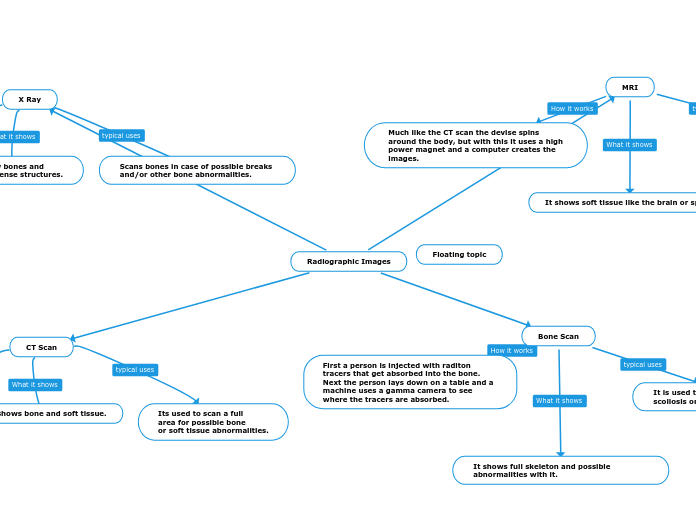
It is used to diagnose bone diseases like scoliosis or breaks.
It shows full skeleton and possible abnormalities with it.
First a person is injected with raditon tracers that get absorbed into the bone. Next the person lays down on a table and a machine uses a gamma camera to see where the tracers are absorbed.
MRI
It is used to check for internal bleeding, brain damage or joint damage.
It shows soft tissue like the brain or spine.
Much like the CT scan the devise spins around the body, but with this it uses a high power magnet and a computer creates the images.
CT Scan
Its used to scan a full
area for possible bone
or soft tissue abnormalities.
It shows bone and soft tissue.
A CT scan is a bunch of
x-rays that take pictures
from multiple angles making
cross section pictures.
X Ray
Scans bones in case of possible breaks
and/or other bone abnormalities.
It show bones and
other dense structures.
They send radiation through
The body and the bones reflect
the radiation making it visible
as white on x-ray pictures.