Categorieën: Alle - functions - neurotransmitters - brain - neurons

door Ema Kralik 5 jaren geleden

740

Chapter 3 - Biological Psychology LECTURE 3

The document delves into the intricate mechanisms and structures that comprise the brain and its functions. It touches upon various parts of the brain, including the hippocampus, temporal lobe, and occipital lobe, each responsible for different functions such as visual processing, memory, and emotion regulation.

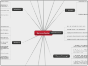
Chapter 3 - Biological Psychology LECTURE 3

History (Of Brain Function)

fMRI

Looks at structures of the brain and the functions

Uses change in blood flow to generate images - best non-invasive technique for brain imaging

PET

Looks at brain structure and brain tissue, shows brain activity during specific tasks

Limitations: needs a radioactive tracer to be injected, there is a delay in reading (40 secs) and it does not shoe which specific area the activity is occuring

MRI

Looks at brain structure and brain tissue

Yields a more 3D image

CAT/CT Scans

Look at brain structure only

Used for quick assessment

335 BCE: Aristotle

387 BCE: Plato

460 BCE: Hippocrates

Middle of 19th Century: Started to really understand the importance of brain

2500 BCE: Trepanation

1700 BCE: Egyptian Mummification

Neo-Cortex

Left Hemisphere: controls academic, verbal and analytical functions

Right Hemisphere: controls creative, artistic and emotional functions

**CORPUS CALLOSUM: connects the left and right brain hemispheres

-receives visual information

-processes auditory info, language and auditory memories

-integrates touch and vision for spacial awareness and orientation

-motor planning, language, memory (complex brain functions and decision making)

Occipital Lobe

Temporal Lobe

Parietal Lobe

Frontal Lobe

The structures and functions of the parts of the limbic system

-memory for spatial locations

-regulates fear and arousal

-endocrine oversees ANS (homeostasis) along with the pituitary glans

-conveys sensory information to the cortex

Hippocampus

Amygdala

Hypothalamus

Thalamus

The structures and functions of the parts of the hind brain

-responsible for coordinating movement and balance (4 F's)

Cerebellum

-responsible for processing vision, hearing and eye movement

-acts as the bridge between the lower and upper ends, responsible for motor control and sensory analysis

-responsible for basic bodily functions such as heartbeat and respiration

Brain Stem

Pons (center end)

Mid Brain (upper end)

Medulla Oblongata (lower end)

Limbic System

Hind Brain

Glutamate: always carry excitory messages for learning and memory

Endorphins: pleasure sensation which controls pain

Peptide Neurotransmittor

Amine Neurotransmittor

Amino Acid Neurotransmittor

Synapses (lock and key): How two neurons communicate and connect with one another to release one or more neurotransmitters

Chapter 3 - Biological Psychology LECTURE 3

Brain Imaging Techniques

The Brain

Carebral Cortex

Neural Cells

Major Neurotransmitters
Adrenaline: involved in stress response
Aspartate: always carry excitory messages
Glycine: always carry inhibitory messages
GABA: most prevelant inhibitory neurotransmittor in CNS
Acetylcholine: used by efferent neurons for memory and learning
Nonephrine: used in ANS in almost every region of the brain (fight-or-flight)
Seratonin: regulates sleep, dreams, mood, pain, appetite, sexual behaviour
Dopamine: produces pleasure sensation
The structures that make up the neural calls
c) Axons and Axon Terminal -axons: portion of neuron that sends signals to other neurons (transmitters) -synaptic vesicle: sac containing neurotransmitters that travels the length of the axon towards the terminal
b) Dendrites -branch-like portion of neuron that receives signals -act as receptors for signals traveling from other neurons, carrying information toward the main body of the nerve cell
a) Cell Body (Soma) -the body of the nerve cell. -manufactures new cell components -contains nucleus