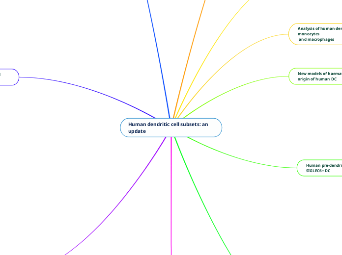
Human dendritic cell subsets: an update
Monocyte-derived inflammatory DC
Functions in immunity
During inflammation, monocyte-derived
cells expand resident populations many-fold.
function mainly at a site of inflammation
Development
DC differentiation is supported by the expression of CD1c, CD1a, FceR1, IRF4, and ZBTB46.
Monocyte-derived cells dominate inflammatory populations, but DC lineage cells may also be recruited in inflammation.
Populations of monocyte-derived cells exist in human steady-state tissues, including the skin, lung and intestine.
Phenotype
Monocytes express CD11c and MHC class II, which are not helpful in separating monocytes from DC.
Inflammatory monocytes retain expression of several markers, including CD13, CD33, CD11b, CD11c, and CD172a.
Inflammatory mo-DC express CD206 and CD209 but also retain expression of CD14 and co-expression of CD16, CD163, and FXIIIA.
CD16+ non-classical monocytes, SLAN+ DC and DC4
DC4
A population of CD16+ cells among HLA-DR+ lineage-negative CD14-negative cells
SLAN expression identifies a subpopulation with lower CD11b, CD14 and CD36 but higher expression of CD16.
SLAN+ cells have monocytic gene expression and recent experiments support the hypothesis that non-classical monocytes differentiate from classical monocytes.
DC4 and SLAN+ cells are likely identical, but their non-redundant roles in immunity are still unclear and further studies are needed.
CD16+ monocytes are heterogeneous and express SLAN, a carbohydrate modification of P-selectin glycoprotein ligand 1.
IRF4/KLF4/NOTCH2-dependent myeloid cDC2
Subsets of cDC2 specializing in Th2 or Th17 responses are dependent upon IRF4 or KLF4, respectively, but this has not been corroborated in humans.
Subsets of cDC2 defined by CD5 and other markers differ in their production of TNF-a, IL-6, IL-10, and IL-23 in response to TLR ligation.
‘Monocyte-like’ cDC2 with lower CD5 are less active in proliferation assays and produce mainly Th1 cells.
CD5-high ‘DC-like’ cDC2 are more active in CCR7-dependent migration, stimulate high naive T-cell proliferation, and preferential priming of Th2, Th17, Th22, and regulatory T cells.
Human cDC2 secrete IL-23, IL-1, TNF-a, IL-8, and IL-10, but are consistently low in the secretion of type III interferon.
Human cDC2 respond well to lipopolysaccharide, flagellin, poly IC, and R848.
Human cDC2 can become high producers of IL-12 and excellent cross-presenting cells
Myeloid cDC2 express a range of lectins, TLRs, NOD-like receptors, and RIG-I-like receptors, similar to monocytes.
TLRs 2, 4, 5, 6, and 8 are present, with notable expression of NOD2, NLRP1, NLRP3, and NAIP.
Transcription factor zbtb46 is not required for the development of cDC2, but is up-regulated during differentiation and also appears on mo-DC.
Myeloid cDC2 require several transcription factors for their development, including GATA2, PU.1, GFI1, ID2, ZEB2, RELB, IRF4, NOTCH2, and KLF4
Heterozygous GATA2 deficiency leads to eventual loss of all cDC2, while bi-allelic IRF8 deficiency also abrogates cDC2 development in humans.
Two subsets of cDC2 have been characterized in human blood, one ‘DC-like’ and the other more ‘monocyte-like’.
Tissue cDC2 spontaneously express low langerin, unlike mouse cDC1, which express langerin.
Myeloid cDC2 are the major population of cDC in human blood, tissues, and lymphoid organs.
CD14+ CD1c+ cells may converge towards the monocyte-like cDC2 phenotype.
CLEC10A (CD301a), VEGFA and FCGR2A (CD32A) as consistent cDC2 markers.
They express CD1c, CD2, FceR1, SIRPA, CD11b, CD11c, CD13, and CD33
Plasmacytoid dendritic cells
Phenotype and distribution
pDCs can prime CD4 T cells, but conflicting roles have been reported in allergy, and tolerogenic pDCs have been proposed to contribute to tumor progression.
Deficiencies in MyD88, IRAK4, and DOCK8 can affect pDC function and number
Specialized in sensing and responding to viral infections
production of tumor necrosis factor and IL-6 is dependent upon the nuclear factor-jB pathway
Sphingosine-1-phosphate signaling interacts with IFNAR to terminate the IFN-a response
FceR1, ILT7, and CD303 (BDCA2) inhibit IFN-a production
IRF7 is the major transducer of type I interferon production in pDCs
Sphingosine-1-phosphate signaling interacts with ILT7 to limit interferon production
CD300A enhances interferon production
Dendritic cell development in mammals is regulated by several transcription factors
GATA2, PU.1, GFI1, IKZF1, and IRF8.
A small subset of CD123+ pDC express CD2+, CD56+, or CD5.
Distinct gene expression pattern that overlaps with myeloid cDC and contain AXL+ SIGLEC 6+ myeloid pre-cDC.
Surface receptors involved in the production of type I interferon
CD303 (CLEC4C; BDCA-2), CD304 (neuropilin; BDCA-4) CD85k (ILT3), CD85g (ILT7), FceR1, BTLA, DR6 (TNFRSF21/CD358), CD300A, FAM129C, CUX2, and GZMB.
pDC retain expression of the GMDP markers CD123 (IL-3R) and CD45RA
pDC do not express myeloid antigens CD11c, CD33, CD11b, or CD13.
Eccentric nucleus and prominent endoplasmic reticulum and golgi.
Langerhans cells
Functions and roles in immunity
Langerhans cells have functional cross-presentation capacity and high MHC class I-related gene expression
LC are capable of local self-renewal, independently of the bone marrow.
Recent lineage tracing experiments in mice indicate that LC have an equal claim to DC and macrophage heritage by virtue of unique dual expression of ZBTB46 and MAFB.
LC are classified as 'macrophages', but they also function as myeloid dendritic cells (DC) that capture antigen, mature, and migrate to lymph nodes.
Langerhans cells (LC) have a shared evolutionary origin with tissue macrophages and microglia of the brain.
Distribution
Langerhans cells migrate to skin-draining lymph nodes and express langerin, CD1a, and CD1c in the T-cell areas
reside in the basal epidermis and other stratified squamous epithelia
They have lower expression of CD11c, CD11b, and CD13 than myeloid cDC2, which can help differentiate between the two cell types
They express langerin and CD1a, FceR1 and CD39
Human pre-dendritic cells and AXL+
SIGLEC6+ DC
Another DC-restricted precursor in human blood described as CD34+ CD100+ with approximately equal cDC1 and cDC2 potential in vitro and the highest enrichment of pre-cDC1 potential in human blood so far described.
This cell has lower expression of CD123 than AXL+ SIGLEC 6+ pre-cDC and appears to be more primitive, owing to its lack of CD116 (GM-CSF receptor) and higher proliferative capacity. It has approximately equal cDC1 and cDC2 potential in vitro and has the highest enrichment of pre-cDC1 potential in human blood so far described.
AXL and SIGLEC 6 emerge as particularly useful markers for a new ‘AS’ DC subset.
A cell population containing AXL+ SIGLEC6+ cells primarily can be classified as precursors, describing them as ‘early pre-DC’ with the ability to develop into cDC1 and cDC2.
The advantage of these recent studies is that they did not begin by excluding mature DC and so found many pre-DC among the CD123+ populations that had been excluded as mature pDC.
CD123 has long been used as a marker to identify pDC but new studies emphasize that myeloid cDC-like cells are also captured in CD123+ populations.
More numerous populations that fit the definition of pre-DC can be found by examining every lineage-negative HLA-DR-positive cell using single-cell transcriptomics or a combination of deep phenotyping and single-cell transcriptomics.
Potential pre-DCs that are DC-restricted precursors that do not yet express the full phenotype of mature DC.
Several groups have reported human pre-DCs that fulfill these criteria, but it is not yet completely clear how they relate to one another.
New models of haematopoiesis and the origin of human DC
DC can be ordered in a spectrum of
phenotypes
myelo-monocytic
lymphoid
DC are a product of the core lympho-myeloid pathway
Analysis of human dendritic cells, monocytes
and macrophages
Transcriptomics
Novel surface markers
Oligonucleotide tags
Combine antibody-based phenotyping with transcriptomics
Fluorescence flow cytometry
Computational tools
Flow-SOM, tSNE, oneSENSE and ISOMAP
"Deep phenotyping"
30–40 antigens
The unified classification of mammalian DC
Robust classification
of DC based primarily on lineage
IRF8 & IRF4
Plasmacytoid DC (pDC)
Conventional’ or ‘classical’ DC (cDC)
CD1c+ myeloid cDC2
CD2, FceR1 and SIRPA
CD141+ myeloid cDC1
CLEC9A, CADM1, BTLA and CD26
IRF8/BATF3-dependent myeloid cDC1
Functions and role in immunity
Human cDC1 possess conserved mechanisms for recognition of viral and intracellular antigens, transport of antigen to endosomal compartments, and production of type III interferon.
Myeloid cDC1 express TLR3, TLR9, and TLR10, with TLR3 playing a role in recognition of dsRNA and production of type I interferons via IRF3.
TLR10 is a negative regulator of TLR signaling and is selectively expressed by cDC1 in humans.
Myeloid cDC1 are a subset of DC with high capacity for cross-presentation of antigens and promotion of Th1 and natural killer responses through IL-12.
Development
Myeloid cDC1 development is dependent upon several transcription factors, including GATA2, PU.1, GFI1, Id2, IRF8, and BATF3
Distribution
Human cDC1 are found in blood and various tissues including lymph nodes, tonsils, spleen, bone marrow, skin, lung, intestine, and liver
Unopposed expression of IRF8 (without IRF4) defines the lineage, but intracellular staining may limit subsequent tests
They are present at approximately one-tenth the frequency of cDC2 in steady-state blood and tissues
Phenotype
CLEC9A, CADM1, BTLA, and XCR1, increase accuracy of identification
They express CD13 and CD33, but differ from cDC2 by low CD11c and little CD11b or SIRPa expression
Human myeloid cDC1 are a subset of blood DC with high expression of CD141+


