Sites where Transcription Occurs
Transcription in Eukaryotes
In nucleus
In a Eukaryotic promoter there is a TATA box: A nucleotide sequence that contains approximately 25 TATA nucleotides upstream from the starting point in the promoter.
Transcription factors (proteins) recognize the TATA box and bind to the DNA strand prior to the RNA polymerase II binds.
Additional transcription factors bind along to the RNA polymerase II.
Transcription in Prokaryotes
In cytoplasm
In Prokaryotes, the RNA polymerase recognizes where to start.
RNA polymerase binds to the promoter (upstream)of a gene, unwinding the DNA to use the strand going from 3' to 5' as the template strand.
#1 INITIATION: RNA polymerase initiates RNA synthesis at the starting point on the template strand.
#2 ELONGATION: The RNA polymerase moves downstream towards transcription, unwinding the DNA and elongating the RNA transcript in a 5' to 3' direction.
#3 TERMINATION: RNA transcript is released and the RNA polymerase detaches from the DNA.
In Eukaryotes the RNA polymerase II transcribes a polyadenylation signal sequence.
This specifies a polyadenylation signal (AAUAAA) into the pre-mRNA bounding it by specific proteins in the nucleus.
AAUAAA cut the RNA transcript free from the polymerase and releases the pre-mRNA.
RNA Processing
The ends of the pre-mRNA are modified. The 5' end is synthesized first and receives a 5' cap. The 5' cap is a modified form of a guanine nucleotide.
RNA splicing
Every 5' end and 3' have UTRs which are untranslated regions. UTRs are parts of the mRNA, but they are not translated into protein- have other functions (ribosome binding).
Spliceosome: a large complex of proteins and small RNAs removes the introns by binding to short nucleotide sequences alone an intron. Then, it releases the intron, and the spliceosome joins the two exons.
A mRNA molecule is formed
mRNA moves from the nucleus to the cytoplasm by passing through the nuclear membrane through a nuclear pore.
Translation occurs
#1 INITIATION:
In Prokaryotes, the small ribosomal subunit binds the mRNA and the tRNA carries fmet as oppose to met.
In Eukaryotes, the small ribosomal subunit binds the mRNA and a specific initiator tRNA carrying the AUG starting codon (methionine).
the small subunit with the tRNA bound, binds to the 5' cap of the mRNA and moves downstream along the mRNA until it reaches the start codon.
Initiator factors (proteins) attach the large ribosomal subunit together, by expending energy obtained by hydrolysis of a GTP molecule.
The initiator tRNA is in the P site (peptidyl-tRNA binding site) while the A site (aminoacyl-tRNA binding site) is ready for the next aminoacyl tRNA. This starts from the N-terminus towards the carboxyl end, C-terminus.
#2 ELONGATION: Amino acids are added to the previous amino acid at the C-terminus end of the growing chain.
A) Codon recognition: The anticodon of an incoming aminoacyl tRNA pairs with the complimentary mRNA codon in the A site. Hydrolysis of GTP releases the accuracy. An appropriate anticodon in the tRNA will bind to the mRNA.
B) Peptide bond formation: An rRNA molecule in the large ribosomal subunit catalyzes the formation of a peptide bond between the amino acids formed in the A site and a growing polypeptide in theP site. The polypeptide is then removed from the tRNA in the P site and is attached to the A site.
C) Translocation: The ribosome translates the rRNA in the A site to the P site. Simultaneously the tRNA in the P site is moved to the E site and it is released. The mRNA moves, and the next codon is translated.
#3 TERMINATION
A) A ribosome reaches a stop codon on the mRNA (UAG, UAA, or UGA) the release factor (shaped like tRNA0 becomes placed in the A site.
B) Hydrolysis is promoted to detach the amino acid of the polypeptide and the tRNA from the P site.
C) The hydrolysis of two GTP molecules dissociate the two ribosomal subunits.
Proteins are made
Codons: mRNA nucleotide triples written in the 5' to 3' direction.
There are regions that are expressed and tare translated into amino acid sequences (Exons)
There are noncoding segments of nucleic acid in between coding regions (Introns)
Functions as a ribozyme which catalyzes its excision
This exports the mature mRNA from the nucleus, protects the mRNA from degradation by hydrolytic enzymes, and help ribosomes attache to the 5' end when it reaches the cytoplasm.
Polyadenylate polymerase adds 50-250 Adenine nucleotides to the 3' end of the pre-mRNA to make a poly-A tail.
In Prokaryotes a RNA sequence called the terminator signals the end of transcription.
While transcription is occurring, DNA strands are re-forming a double helix.
RNA polymerase moves along the DNA untwisting the double helix and exposing approximately 10-20 DNA nucleotides at a time to pair it with RNA nucleotides.
Direction of transcription = downstream The other direction = upstream
Transcription Initiation Complex: when transcription factors and RNA polymerase become bound to the promoter.
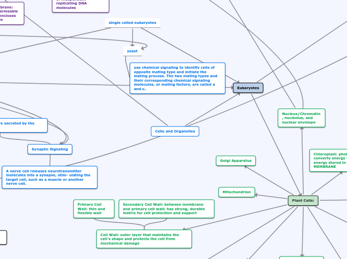
single celled eukaryotes
yeast
use chemical signaling to identify cells of opposite mating type and initiate the mating process. The two mating types and their corresponding chemical signaling molecules, or mating factors, are called a and 𝛂.
Cell Signaling
Local Signaling
Signaling molecules are secreted by the signaling cell
Synaptic Signaling
A nerve cell releases neurotransmitter molecules into a synapse, stim- ulating the target cell, such as a muscle or another nerve cell.
Paracrine Signaling
In animal cells, a signaling cell acts on nearby target cells by secreting molecules of a local regulator
Communication by direct contact between cells can happen through cell junctions or cell-cell recognition
cell-cell recognition: Two cells in an animal may communicate by interaction between molecules protruding from their surfaces.
cell junctions: Both animals and plants have cell junctions that allow molecules, including signaling molecules, to pass readily between adjacent cells without crossing plasma membranes.
plants have plasmodesmata
animals have gap junctions
Regulation of Gene Expression
eukaryotic gene expression is regulated at many stages
Differential Gene Expression: expression of different genes by cells with the same genome
Processes most often regulated are: Chromatin modification, transcription, rna processing, transport to cytoplasm, translation, protein processing, protein processing and degradation and transport to cellular destination
regulation of transcription initiation: control elements in enhancers bind specific transcription factors, bending of the DNA enables activators to contact proteins at the promoter, initiating transcription
Translation: initiation of translation can be controlled via regulation of initiation factors
RNA processing
alternative RNA splicing: results in a single gene coding for multiple proteins
Chromatin modification:
dan methylation: reduces transcription
histone acetylation:loosens chromatin structure, enhancing transcription
initiation of translation can be controlled via regulation of initiation factors
Cells control metabolism by regulating the expression of genes coding for
enzymes
Bacteria: genes are clustered into operons and have an operator site on the DNA which switches the operon on or off
Positive Gene Regulation
Some operons are also subject to positive gene regulation via a stimulatory activator protein, such as cAMP receptor protein (CRP), which, when activated by cyclic AMP, binds to a site within the promoter and stimulates transcription.
Repressible and Inducible operons are examples of negative gene regulation
Repressible:the repressor is active when bound to a corepressor, usually the end product of an anabolic pathway.
lac operon: repressible
lactose absent
operon off since repressor binds to operon
Lactose present
repressor bound to lactose
no glucose
adenyl cyclase active, cAMP levels high
CAP active
operon on
glucose present
inactive adenyl cyclase, cAMP levels low
CAP inactive
operon off
Inducible:binding of an inducer to an innately active repressor inactivates the repressor and turns on transcription. Inducible enzymes usually function in catabolic pathways.
Trp operon is usually on (inducible) but can be turned off.
When a trphlophan molecule(corepressor) binds to trp repressor at an allosteric site,
the repressor protein becomes active
attaches to operon
turns operon off
Leslie Oros (Transcription & Translation)
Jaqueline Valdez (Cells and Organelles & DNA Replication)
Jasmine Serna (Cell Signaling & Gene Regulation)
Some pathways lead to a nuclear response: Specific genes are turned on or off by activated transcription factors. In others,
the response involves cytoplasmic regulation.
In both Prokaryotic and Eukaryotic cells, a ribosome brings mRNA and together with tRNA carrying amino acids. It is divided by a small subunit, where the mRNA binds, and a large subunit where it has three binding sites for tRNA. The order from left to right is E site, P site, A site.
tRNA (transfer RNA): translates the message in a series of codons along an mRNA molecule.
Structure: A two-dimensional structure has four base-paired regions and three loops. The base sequence of the amino acid is attached at the 3' end. The anticodon triplet and some sequences on the other loops are different for each tRNA. A three-dimensional structure has hydrogen bonds that has the amino acid attachment site on one end and the anticodon at the bottom from 3' to 5'
Anticodon: a nucleotide triplet that base-pairs to a specific mRNA codon.
3 Stages of Cell Signaling
2. Transduction: the binding of the signaling molecule changes the receptor protein In some way, initiating transduction. The transduction stage converts the signal to a form that can bring about a specific cellular response
3. Reception:the transduced signal finally triggers a specific cellular response
1.Reception: the target cell detects the signaling molecule coming from outside the cell. The signal is detected when the signaling molecule binds to a receptor protein located at the cell’s surface
Long-Distance signaling
Both plants and animals use hormones for long distance signaling
Plant hormones (often called plant growth regulators) sometimes travel in plant vessels (tubes) but more often reach their targets by moving through cells or by diffusing through the air as a gas
Endocrine (hormonal) signaling: Specialized endocrine cells secrete hormones into body fluids, often blood. Hormones reach most body cells, but are bound by and affect only some cell
Receptors in the plasma membrane:Most water-soluble signaling molecules bind to specific sites on transmembrane receptor proteins that transmit information
ion channel receptors
1.When the ligand binds to the receptor and the channel opens, specific ions can flow through the channel and rapidly change the concentration of that particular ion inside the cell.
2, When the ligand dissociates from this receptor, the channel closes and ions no longer enter the cell.
receptor tyrosine kinases
1. the receptor tyrosine kinase first exist as individual monomers
2. the binding of a signaling molecule causes 2 receptor monomers to associate closely with each other forming a dimer
3. Dimerization activates the tyrosine kinase region and each tk adds a phosphate from atp to a tyrosine that is part of the tail of the other monomer
4. receptor is fully activated, it is recognize by specific relay proteins within the cell
G protein-coupled receptor
Works with the help of a G protein, a pro- tein that binds the energy-rich molecule GTP.yeast mating factors, neurotransmitters, and epinephrine (adrenaline) and many other hormones—use GPCRs.
!. GTP binds to the G protein, while it is inactive and the protein & receptor work together with an enzyme.
2. The signaling molecule binds to the extracellular side of the receptor, causing it to change shape, and GTP displaces GDP,, protein activated
Actiavted G protein binds to an enzyme altering its shape and activity and can now trigger a cellular response
GTPase function of the G protein allows the pathway to shut down rapidly when the signaling molecule is no longer present.
Cells and Organelles
Eukaryotes
Animal Cells:
Endoplasmic Reticulum (ER): a network of membranous sacs; the ER membrane separates the internal compartment of the ER
Smooth ER: outer surface, lacks ribosomes, functions in metabolic processes (synthesis of lipids, metabolism of carbohydrates, detoxifies drugs and poisons, breaks down hormones
Rough ER: studded with ribosomes on the outer surface of the membrane, makes secretory proteins
Nucleus: contains most of the DNA; DOUBLE MEMBRANE
Chromatin: material consisting of DNA and proteins; visible in cell division as individual condensed chromosomes
Nucleolus: nonmembranous structure involved in production of ribosomes
Nuclear envelope: DOUBLE MEMBRANE enclosing the nucleus, separating its contents from the cytoplasm, pierce by nuclear pores that serve as "holes" that regulate entry and exit of materials through envelope; continuous with ER
Ribosomes: complexes that synthesize proteins; NO MEMBRANE
Bound Ribosomes: attached to the outside of the ER or nuclear envelope & make proteins for insertion into membranes
Free Ribosomes: suspended in the cytosol, proteins of these function within cytosol
Golgi Apparatus: organelle active in synthesis, modification, sorting, and secretion of cell products
Trans face: shipping department of Golgi, gives rise to vesicles that pinch off and travel to other sites
Cis face: receiving department of Golgi, located near the ER, transport vesicles used to more from ER to Golgi, ER lumen and membrane fuse with the Golgi membrane
products of the ER are modified, stored, and then sent to other destinations
Extracellular Matrix: helps cells to bind together and regulates several cell functions; made out of glycoproteins and carbohydrates.
Cells attach to the matrix with the help of fibronectin- binds to cell-surface receptor proteins (integrins) in the membrane
Contains collagen (glycoprotein) that forms strong fibers outside the cells
Centrosome: region where the cell's microtubules are initiates; contains a pair of centrioles
Flagellum: motility structure present in some animal cells; composed of microtubules
Cytoskeleton: reinforces cell shape, functions in cell movement, and components are made of protein; located in cytoplasm
Microfilaments: thin solid rods that are built from actin molecules; help maintain cell shape, changes in cell shape, muscle contraction, cytoplasmic streaming, cell motility, and cell division
Actin-protein interactions help cytoplasmic streaming (the circular flow of cytoplasm within cell)
Actin filaments and Myosin (motor protein) interact to cause contraction of muscle cells
Intermediate filaments: maintain cell shape, anchorage of organelles, and help with formation of nuclear lamina
Microtubules: hollow rods constructed from globular proteins called tubulins; maintain cell shape, cell motility in cilia and flagella, chromosome movements in cell division, and organelle movements
Dyneins have two "feet" that "walk" along the micotubules and use ATP to cause bending movement
Large motor proteins called dyneins are attached along each outer microtubule double and are responsible for the bending movement of flagella and cilia
Microvilli: projections that increase the cell's surface area
Peroxisomes: produces hydrogen peroxide as a by-product and then converts it to water; single-membrane organelle
Mitochondrion: organelle where cellular respiration occurs and most ATP is generated; double-membrane organelle
Inner membrane: coiled with infoldings (cristae) and divides the mitochondrion into TWO internal compartments
Mitochondrial Matrix: enclosed by the inner membrane, contains different enzymes and mitochondrial DNA and ribosomes
Intermembrane Space: narrow region between the inner and outer membrane
Outer membrane is SMOOTH
Lysosomes: digestive organelle where macromolecules are hydrolyzed (add water molecule); has hydrolytic enzymes and are single-membrane organelles
Autophagy: lysosomes fuse with vesicle containing damaged organelles and hydrolytic enzymes are used to recycle the cell's own organic material
Phagocytosis: lysosomes fuse with a food vacuole and hydrolytic enzymes digest the food particles
excessive leakage from A LOT of lysosomes leads to self-digestion of cell
Plant Cells:
Ribosomes
Rough and Smooth ER
Nucleus/Chromatin, nucleolus, and nuclear envelope
Sites where DNA Replication Occurs
Each cell has an ORI (origin of replication) where DNA replication begins; prokaryotes have one ORI and eukaryotes have many
1) Helicase: enzyme that untwists the 2 strands at the replication fork
2) Single-stranded Binding Proteins: bind to the unpaired DNA strands and keep them from re-pairing
3) Primase: enzyme that synthesizes a complementary RNA primer using the parent strand as the template; new DNA strand will start at the 3' end of the primer
4) DNA Polymerase III: enzyme that adds a DNA nucleotide to the 3' end of the primer and continues to add complementary DNA nucleotides to the template strand
5) DNA Polymerase I: removes RNA nucleotides of primer from 5' end and replaces them with DNA nucleotides
6) DNA Ligase: joins final nucleotide of DNA replacement to the first DNA nucleotide of the adjacent Okazaki fragment
Nuclease: a DNA-cutting enzyme that cuts a segment of DNA strand that contains damage; the gaps are filled with nucleotides using undamaged strand
Sliding clamp: converts DNA Poly. III from being distributive (falling off) to processive (staying on)
Replicates the leading strand 5' to 3' direction; continously; only one primer
Replicates the lagging strand away from replication fork, 5' to 3' direction, and discontinously synthesized in a series of Okazaki fragments, who each need a primer
Adds each monomer through dehydration reactions; is able to proofread its own work
Needs a primer & can only replicate 5' to 3' end
Topoisomerase: enzyme that helps relieve the strain by breaking, swiveling, and rejoining DNA strands
Golgi Apparatus
Mitochondrion
Peroxisomes
Cytoskeleton: only microfilaments and microtubules
Central Vacuole: functions include storage, breakdown of waste products, hydrolyze macromolecules, and plant growth
Plasmodesmata: cytoplasmic channels through cell walls that connect the cytoplasms of adjacent cells
Chloroplast: photosynthetic organelle; converts energy of sunlight to chemical energy stored in sugar molecules; DOUBLE MEMBRANE
Stroma: fluid outside the thylakoid, contains the chloroplast DNA, ribosomes, and enzymes
Membranes of the chloroplast divide the chloroplast into 3 compartments: intermembrane space, stroma, and thylakoid space
Thylakoids: another membranous system inside chloroplasts in the form of flattened, interconnected sacs; thylakoids are stacked; each stack is called a granum
Cell Wall: outer layer that maintains the cell's shape and protects the cell from mechanical damage
Secondary Cell Wall: between membrane and primary cell wall; has strong, durable matrix for cell protection and support
Primary Cell Wall: thin and flexible wall
Prokaryotes
Cytoplasm: interior of the cell
Cytosol: semifluid, jelly-like substance where cellular components are suspended
Peptidoglycan: located in the cell walls of BACTERIA CELLS and provides rigid mechanical support and anchors molecules
Endospores: help the cell survive under harsh environmental conditions i.e. lacking water or essential nutrients
Pili: appendages that help attach 2 cells together and used for bacterial mating
Flagella: organelle used for movement
Glycocalyx: outer coating of many prokaryotes, consisting of a capsule or slime layer
Slime layer: unorganized layer of extracellular fluid that protects the cell against dehydration (and other dangers) and helps it adhere to surfaces
Capsule: sticky layer of polysaccharides or proteins that protects the cell, helps maintain moisture, and helps the cell adhere to surfaces
Plasma membrane: selectively permeable barrier that encloses the cytoplasm
Ribosomes: complexes that synthesize proteins
Nucleoid: region where DNA is at (NO MEMBRANE); have a circular chromosome
Plasmids: smaller rings of independent replicating DNA molecules
Fimbrie: attachment structures on the surface of some prokaryotes; also used for bacterial mating
Cell wall: rigid structure outside of the plasma membrane that gives the cell shape
