Категории: Все - mri - bones - imaging

по Emma Sanford [STUDENT] 2 лет назад

90

Radiographic Images

Radiographic imaging techniques such as X-rays, CT scans, and MRIs are essential tools in modern medicine. X-rays utilize photons to capture images of the internal structures of the body, primarily showing bones and identifying fractures or diseases like bone cancer.

Radiographic Images

Radiographic Images

Bone Scan

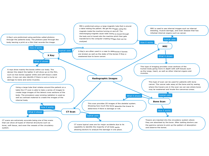
This type of scan can be used for patients with bone cancer. The cancer eats away at the bone and by seeing where the tracers are in the scan we can see where bone may be weakened and locate the cancerous areas.
This scan provides 2D images of the skeletal system. Showing how much the bone absorbs the tracer to determine if there is damage or not.
Tracers are injected into the circulatory system where they are absorbed by the bone. After waiting doctors use the gamma camera to pick up the pattern of absorption and observe the bones.

MRI

MRI is used to see internal images such as internal bleeding, muscle damage, and even disease that has infected internal organs such as cancer.
This type of imaging provides cross sections of the human body going more in depth with soft tissues such as the lungs, heart, as well as other internal organs and muscles.
MRI is preformed using a a large magnetic tube that is around a table holding the patient. We get the images using the magnets inside the machine turning on and off. The interchanging magnets cause radio waves to travel through the body and is traced onto the machine which then gets transferred to the computer creating images that can be analyzed.

CT Scan

CT scans typical uses are for major accidents due to its capability to identify the majority of all body parts allowing doctors to analyze the damage in one place.
CT scans are extremely accurate being one of few scans that can show all types of internal structures such as bone, soft tissue, and even the vessels of the circulatory system.
Using a large tube that rotates around the patient on a table the CT scan is able to take a series of images to create large images of the lateral cross sections of the body. The procedure uses ionizing radiation is used as well as contrast material to create the images of the internal body.

X Ray

X-Ray's are often used in a case to determine if bones are broken as well as the state of the bones if they a weakened due to bone cancer.
X-rays show mainly the bones within our body. The denser the object the lighter it will show up on the film, such as how bones appear white and soft tissue a dark grey. X-rays can also identify if there is such a lump or damage to bone and some muscles.
X-Ray's are preformed using particles called photons through the patients body. The photons pass through the body leaving a print on a film that records the image.