TAB makes a new subtopic
To make a new line in a topic box
hit "shift+enter"
TIPS and TRICKS
To move a subtopic to be under a new topic
just drag and drop it on top of the topic
that you want it to be associated with
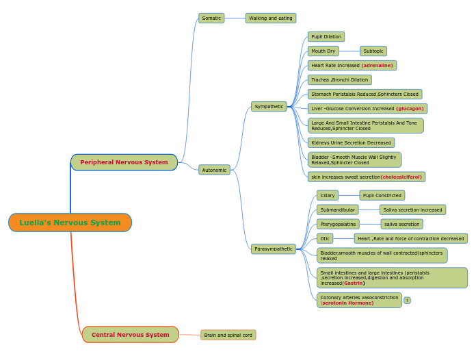
Autonomic Nervous System
The classic definition of the ANS is the preganglionic and postganglionic neurons within the sympathetic and parasympathetic divisions.
A modern definition of the ANS takes into account the descending pathways from several forebrain and brainstem regions as well as visceral afferent pathways that set the level of activity in sympathetic and parasympathetic nerves.
Enteric
Sympathetic
ventral root
white rami communicantans
sympathetic paravertebral ganglion
Paravertebral ganglia are located adjacent to each thoracic and upper lumbar spinal segment; in addition, there are a few ganglia adjacent to the cervical and sacral spinal segments. The ganglia are connected to each other via the axons of preganglionic neurons that travel rostrally or caudally to terminate on postganglionic neurons located at some distance. Together these ganglia and axons form the sympathetic chain bilaterally. This arrangement is seen inFigure 13–2 and Figure 13–3.
PRE-VERTEBRAL GANGLIA
Some preganglionic neurons pass through the paravertebral ganglion chain and end on postganglionic neurons located in prevertebral (orcollateral) ganglia close to the viscera, including the celiac, superior mesenteric, and inferior mesenteric ganglia (Figure 13–3). There are also preganglionic neurons whose axons terminate directly on the effector organ, the adrenal gland.
SYMPATHETIC CHAIN*
gray rami communicans
The axons of some of the postganglionic neurons leave the chain ganglia and reenter the spinal nerves via the gray rami communicans and are distributed to autonomic effectors in the areas supplied by these spinal nerves (Figure 13–2). These postganglionic sympathetic nerves terminate mainly on smooth muscle (eg, blood vessels and hair follicles) and on sweat glands in the limbs. Other postganglionic fibers leave the chain ganglia to enter the thoracic cavity to terminate in visceral organs. Postganglionic fibers from prevertebral ganglia also terminate in visceral targets.
aka " thoracolumbar division of the ANS"
shorter pre-, longer post-ganglionic neuron
Parasympathetic
pelvic nerve
pelvic viscera via branches of the second to fourth sacral spinal nerves
dorsal motor vagal nucleus
esophagus, trachea, lungs, and gastrointestinal tract
nucleus ambiguus
sinoatrial (SA) and atrioventricular (AV) nodes in the heart
Edinger–Westphal nucleus
ciliary ganglia
innervate the sphincter (constrictor) muscle of the iris and the ciliary muscle.
superior salivatory nucleus
inferior salivatory nucleus
otic ganglion
parotid salivary gland
sphenopalatine ganglia
submandibular ganglia
submandibular (also called submaxillary) and sublingual glands
AKA craniosacral division
because of the location of its preganglionic neurons; preganglionic neurons are located in several cranial nerve nuclei (III, VII, IX, and X) and in the IML of the sacral spinal cord
longer pre-, shorter post- ganglionic neuron
Level 3 - stop at this level


