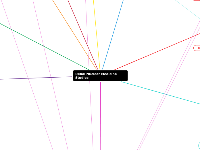
Renal Nuclear Medicine Studies
Vesicoureteral Reflux Scan
Draw ROIs around intrarenal collecting systems and bladder
Calculate the residual urine volume
Formula for calculating the residual volume (mL)= Voided volume (mL) x Residual counts/min
________________________________________________
Max counts/min - Residual counts/min
Abnormal: Activity in upper urinary tract during filling, at full capacity, and /or while voiding.
Normal: No reflux visualized. Nearly all of solution is voided from bladder.
Remove the patients foley catheter. Then measure the urine.
Take an immediate post-void static (120 sec)
Deflate the catheter balloon and have the patient void while taking a dynamic image (2sec/frame for 120sec)
Take a immediate static once the bladder is full (120sec)
If Reflux is seen, record the amount of saline that was infused
Fill the bladder until drip from the bag slows or the patient begins to void around the catheter.
To determine the max capacity of a patients bladder use the formula: (age+2) x 30= Volume instill (mL)
Prepare camera for dynamic 60sec fill. Then inject Tc99m sulfur colloid into the tubing connected to bladder catheter. Begin to fill bladder with saline and begin the dynamic image.
Place the foley catheter using aseptic technique. Inflate the balloon and tape to secure.
Prep 500mL saline bag and the Tc99m sulfur colloid
Prep the patient by having them void or by making sure they are in a clean diaper if an infant.
Views: Posterior, LPO, and RPO with bladder and kidneys on the FOV
Patient Position: Sitting if using the potty seat, or lying supine of the table
Tc99m- Sulfur Colloid
MOL: Compartmental localization with the flow of Urine
Route of Administration: Via Foley catheter tubing
Dose: .5-1mCi
Ancillary Equipment: IV pole and potty seat (if needed)
Post-Void: 120sec static image
Void: Dynamic 2sec/ frame for 120sec.
Data Set: 60 images
Pre-Void: 120sec static image
Data Set: 1 image
Bladder filling: Dynamic 5sec per frame for 60 sec.
Data Set: 12 images
Patient Prep.
Prepare a 500mL bag of saline
Have the patient void before the exam. If the patient is an infant, have them wear a clean diaper.
Obtain a written consent for catheterization
Thoroughly explain the procedure to both the patient and the parents
Place absorbent pads around the potty chair if it is going to be used
Indications: Evaluation and detection of vesicoureteral reflux (VUR)
Morphological Renal Imaging
Additional Patient Prep: If this is a pediatric patient, a parent must sign a informed consent for sedation to begin study
Processing: No processing, it is a qualitative study
Abnormal: To differentiate a Column of Bertin from a true mass, DMSA will show uptake and a mass will not have any uptake. If Acute Pyelonephritis is the issue it may appear as single or multiple defects resulting in decreased uptake.
Normal: A smooth renal contour. Uniform and equal tracer concentration
Inject the Chosen Rph, then wait 2hrs before imaging
Select the proper collimator
Parallel or Pinhole Collimator
- A Pinhole collimator is preferred for children
Place the patient supine and begin taking static images (approximately 3min per view)
A SPECT can also be done (Approximately 15-30min)
Views: Ant, Post, and an optional RAO,LAO,RPO,LPO
Position: Supine
Rph
Tc99m- DMSA or Tc99m- GH
Go to Radiopharmaceutical section for Dose, MOL, and ROA
SPECT: 180 Rotation with 40sec/stop for 40 Views
Statics
Pinhole Collimator: 100K counts per image
Parallel hole colimator: 500K counts per image
Data set:2-6 images
Matrix: 128x128
Energy: 140keV
Confirmation of suspected hypertrophic column of berlin
Adema or scarring from acute pyelonephritis
Renal with ACE
Optional Maneuvers
Some protocols give the patient lasix just like a diuretic scintigraphy to empty any pelvic retention
A baseline renal scintigraphy without ACE should be done either before the Scintigraphy with ACE, or it can be done 2-3 days post Ace renal scintigraphy
Processing: Renogram
Abnormal: Asymmetric curve, low uptake, little to no excretion from kidneys, bladder activity does not increase
Normal: Symmetric curve, prompt kidney uptake, Decreased kidney activity to T1/2 at 8-12min mark, bladder activity increases as the kidneys excrete
Views: Posterior with both kidneys and bladder in the FOV
Patient position: Supine
Get the Baseline BP
Give the patient the chosen ACE inhibitor and wait the designated time
If captopril wait 1 hr. before Rph injection and take BP every 15 min
If Enalaprilat wait 10 min before Rph injection
Inject the chosen Rph and begin the flow immediatly (2min)
Begin the Dynamic immediately after flow (20-30min)
Take the patient final BP
RPh/ Interventional pharmaceuticals
Interventional Pharmaceuticals
Enalaprilat: 40ug/kg
Administered via IV
Wait 10 min before the injection of Rph
Captopril: 25-50 mg
Pill is crushed up and added to water
Administered PO
Must be given 1HR prior to Rph injection
Rph: 5-10 mCi of Tc99m-DTPA or Tc99m-MAG3 (MAG3 is the preferred RPH)
Refer to Radiopharmaceutical section for MOA, and MOL
Number of images
Dynamic: 30sec/frame for 20-30 min
Data set: 40-60 images
Data acquisition
Energy:140 keV
Additional Contraindications
Stop taking ACE inhibitors/diuretics 4 days prior to exam
Patient was not NPO for at least 4 hrs
Obtain a baseline BP and start an IV
NPO 4-6 Hrs before exam
To assess if patients hypertension is related to possible RAS (renal artery stenosis: the narrowing of one or more arteries that carry blood to your kidneys.)
Diuretic Renal Imaging
Additional Patient Prep
Stop taking diuretics 4 days prior to exam
Abnormal: Asymmetric curve, No excretion from the kidneys/ no filling of the bladder, and slow activity increase in kidneys
Normal: Symmetric curve, excretion begins 2-3 min post Lasix injections, and the bladder curve increases post Lasix injection.
Processing
Imaging
Procedure
Bolus Injection of Rph
Immediately begin the flow imaging
Begin Dynamic imaging immediately after the flow. (20min)
Once the dynamic has been going for 20min, Inject the Lasix (40mg for an adult)
Some protocols have the patient void before the Lasix injection.
The Lasix is a slow injection over a 1-2 min period
Continue dynamic for 20min
Have the patient void and then do a post void static. The patient can either be is the supine position or upright with there back to the detector.
Patient Position
Supine
Some protocols may want the patient to be in an upright position with there back to the camera. This is mostly for post void imaging.
Views
Posterior with both kidneys and bladder within the FOV
If the patient has had a renal transplant than images should be acquired in the anterior view
Rph/Interventional pharmaceuticals
Lasix: 40 mg
If a pediatric patient dose is 1mg/kg with a max of 40mg
Tc99m-MAG3 is the preferred Rph
Go to the descriptions of the Rph for dose, MOL, and MOA
Data Analysis
Number of Images
Dynamic: 30 sec/frame for 20 min
Data set: 40 images
Flow: 2sec/frame for 2 min
Matrix
Data Acquisition
Energy: 140 keV
Dynamic/Flow and Static
Evaluate renal function and urodynamics
To distinguish between obstructive hydronephrosis and non-obstructive collecting system dilation
Radiopharmaceuticals
Morphological Radiopharmaceuticals
(AKA cortical agents)
Agents that are rapidly taken up by the kidneys, BUT use more complex mechanisms that involve ERPF, GFR, Tubular secretion, AND tubular resorption
Tc99m- DMSA
AKA: Succimer
Dose:
- Adult: 5mCi
- Pediatric: 55uCi/kg; minimum of 500 uCi
Administered via IV
34% is retained by the renal cortex
MOL: Tubular binding
Tc99m-glucoheptonate
No longer used in the US
5-10% retention by the renal cortex
Dose: 10-15mCi
ROA: IV
MOL: Glomerular filtration and tubular secretion
Functional Radiopharmaceuticals
These are rapidly taken up and excreted by the kidneys by a single, simple physiological mechanism (i.e. ERPF or GFR)
Tubular Agents
Tc99m-MAG3
Dose:
- Adult: 10-20 mCi
- Pediatrics: 100 uCi/kg; minimum 1 mCi
Administered via IV
MOL: binds to RBC
Clearance 100% via tubular secretion
Used to measure ERPF
Recommended in pts with decreased renal function and infants
High first pass ratio
High target to background ratio
Great image quality, low radiation dose
I-131 Hippuran
Dose:
- Adult: .025-.2mCi
- Pediatrics: Not used in pediatrics
Administered via IV
MOL: ion exchange
No longer available in the US due to the high radiation dosage and low image quality
Used to measure ERPF
High first pass extraction: almost all of the radiation delivered to the kidneys remains in the kidneys.
Near total tubal secretion
High target to background ratio
Glomerular Agents
Tc99m-DTPA
Dose:
- Adult: 10-20 mCi
- Pediatric: 200uCi/kg; minimum 2mCi
Administered via IV
MOL: Inulin analog
Excreted 100% via glomerular filtration within 2 hrs.
Assess renal blood flow, function, drainage of the pelvicalyceal systems
Measures GFR
Readily available
Great image quality, low radiation dose to patient
I-125 Iothalamate
Dose:
- Adult: 80 uCi
- Pediatric: 1uCi/kg; minimum 10 uCi
Administered via IV
MOL: simple/passive diffusion
AKA: Glofil
Measurement of GFR
Poor image quality
Contraindications
Patient is unable to remain still
Dehydrated
Currently taking Diuretics
A recent renal arteriogram with contrast
Pregnancy/Breastfeeding
Patient Prep
Detailed Patient History
Do you still have both of your kidneys?
Previous transplant?
Also look in patients records for:
- previous imaging done to see kidney location/size
- creatinine levels (high number may indicate kidney failure)
If the patient has a nephrostomy, check the order to see if the tube should be clamped or not.
Make sure patient has not had a recent renal arteriogram with contrast.
Patient needs to void before scan and those who cannot void bladder need a urinary catheter
Start IV
Adequately hydrated
Oral hydration of 20 ounces (App. 3 cups an Hr. before imaging) or IV hydration of 250 ml prior to images
Equipment and Parameters
IV Supplies
Single or Dual head gamma camera
LEAP collimator
Zoom FOV for pediatrics
Typically a zoom of 2
Full FOV for adults
Basic Renal Scan (Functional renal imaging)
Indications
Evaluation of renal perfusion and function, and collecting system patency information
Interpretation
Abnormal: more uptake in one kidney than the other. A lower excretion rate. Asymmetric curve
Normal: the curve for each kidney should be reasonably symmetric with prompt uptake and a peak between the first 3-5 min. A decreased slope of less than 50% at the 20 min mark.
If MAG3 was used then a ERPF of 600 mL/min is normal
If DTPA was used then a GFR of 120 mL/min is normal
Processing:
Renogram
A renogram is a time-activity curve that provides a graphic representation of the uptake and excretion of a rph.
Draw ROI around the aorta, bladder, both kidneys, and a background below each kidney
This creates the information to make the renogram
Imaging:
Procedure:
Bolus inj.
Immediately begin flow
Begin the dynamic function image after flow
Have the patient void then do the post void image standing with back to detector
Views: Posterior (anterior if patient has had a renal transplant)
Patient Position:
-Supine (can be standing for post void)
-Sternal notch to include both kidneys, inferior bladder in FOV
RPH
Tc99m-MAG3 or Tc99m-DTPA
Look at Rph section for dose amount MOA and MOL
Instrumentation
Data analysis
Number of images:
Static: 2min
Data set: 1
Dynamic: 30sec/frame for 30 min
Data set: 60 images
Flow: 2sec/frame for 1-2min
Data set: 30-60 images
Matrix:
Dynamic: 256x256
Flow: 128x128
Data Acqusition
Energy peak: 140keV
Window: 20%
Dynamic/flow and static


