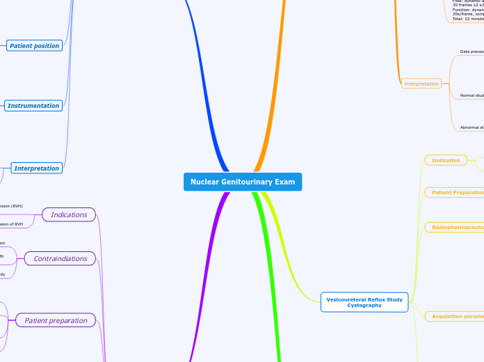
Nuclear Genitourinary Exam
The Personal SWOT Analysis will provide insights based on your personality strengths and weaknesses, challenges you see ahead, opportunities present around you now, as well as future favorable circumstances.
Renal with ACE
Processing
If abnormal, will need a follow-up study without
Captopril for comparison
Generate time-activity renogram curves
Draw 2 bg regions of interest
(one for each kidney)
Draw region of interest around each
kidney and the aorta
Flow study:
2 sec/ frame for 1 min
then stats every 30 sec for 20 min
Views: posterior
LFOV gamma camera
5-10 mCi of Tc99m-MAG3 via IV
Enalaprilat:
40 ug/kg IV over 3-5 min
wait 15 min then inject Rph
Captopril
50 mg pill, PO 1 hr before procedure
Obtain patient baseline of BP
ACE inhibitors and angiotensin II
receptor blocking agents should be
discontinued for 4-7 days
Diuretics should be discontinued for 3 days
Only liquids for 4 hr before procedures
well hydrated
Contraindiations
any recent nuclear medicine study
Breast-feeding should be provided with
appropriate radiation safety
Pregnant must be excluded for this exam
Diagnosis or exclusion of RVH
Differentiate of renal vascular hypertension (RVH)
from renal artery stenosis
Diuretic Renal Scintigraphy
Have high sensitivity for the detection of urodynamically
significant ureteral narrowings.
If a urodynamically significant outflow obstruction is present,
the affected kidney is unable to increase its urine flow rate
in response to the furosemide injection.
Energy: 140 keV
Window: 20%
Matrix: 128x128
Dynamic/flow
Detector system
Collimator: LEHR
camera: LFOV Gamma Camera
Patient position
Use dose to mark sternal notch in top one third of image and bladder in bottom one third of image. Use dose to ensure that patient's left and right sides are in the FOV.
Supine
Furosemide:
20-40 mg for adults
0.5-1 mg/kg for pediatrics
injected slowly over 1-2 min
image is continued for 20 min
ROA: IV bolus
MOL: MAG3 is tubular secretion/
DTPA is glomerular filtration
5 mCi Tc99m-DTPA may also be used
Best with 10 mCi of Tc99m-MAG3
Patient should be well hydrated
drink 10 to 16 oz of water over 30 min
Technologist need to start an IV on patient
Patient does not to need to be NPO
Light meal and adequate hydration
Technologists need to start an IV
Contraindications
Furosemide is contraindicated in anuric/
or dehydrated patients
Pregnant/ breast-feeding:
pregnant must be excluded for this exam
breast-feeding patient use the appropriate
radiation safety
Distinguish between obstructive hydronephrosis
and nonobstructive collecting system dilation
Evaluation of hydronephrosis
Evaluation of renal obstructive nephropathy
Morphological study
Renal cortical scintigraphy
Abnormal study:
congenital abnormalities: horseshoe kidney,
ectopic kidney, and the absence of kidney.
Normal study:
show smooth renal contour
Both kidneys should have equal amount of
radioactivity and uniform tracer distribution
Time/view:
Dynamic: 2-4s / frame for 60-120 seconds
Static: 500k to 1 M counts
SPECT: 2-4 hours post injection. 25-30 s/projections
Injection to imaging time:
immediate and 2-4 hours after injection
24 hour delay is optional
Patient position: supine
camera position: posterior
Camera type: LFOV gamma camera
Energy: 140 keV
Window: 20%
Collimators: LEHR
Matrix: 128x128
Pediatric patient: 50 uCi/kg
15-20 mCi of Tc99m-GH
5 mCi of Tc99m-DMSA
Patient preparation
Subtopic
Void prior to imaging
Well hydrated
detection of the presence of absence of small renal infarctions
Evaluation of renal cortex
Identification of functioning renal tissue in patients
with congenital abnormalities
Differentiation of renal mass from normal variant
Confirmation of suspected column of Bertin
Detection of pyelonephritis
Vesicoureteral Reflux Study
Cystography
Abnormal study:
show ureteral reflux, especially during urination
Reflux bladder volume and the volume of reflux into the kidney can also be calculated
Normal study:
Increasing activity in the bladder without
reflux into the ureters
Total bladder volume of residual postvoid volume
and bladder volume at initiation of reflux can be measured.
Acquisition parameters
Time/view:
dynamic: 10-15 s/frame
static: 120 seconds
Additional views: full bladder
RAO/left anterior
oblique postinfusion
postvoid
Patient position:
Supine
Data acquisition
Collimator: LEHR
Matrix: 128x128
Window: 20%
Energy: 140 keV
Camera type: LFOV gamma camera
Camera position: Posterior
Radiopharmaceutical
ROA: into the Foley catheter
by injection port
1 mCi of Tc99m-sulfur colloid or DTPA
establish an indwelling Foley catheter
Ask patient to void before catheterization
Indication
Commonly perform in children
Evaluation and detection of vesicoureteral reflux
Basic Renal Scan
Begin by identifying your strengths. These are the traits or skills that set you apart from others.
List out all your strengths - if you get stuck, talk to people around you and ask for their input. Please be honest with yourself.
Interpretation
Abnormal study
Areas of relative decreased activity resulting from cysts
or avascular tumors may also be seen in the flow sequence.
Normal study
Activity will be seen in the renal collecting system,
ureters, and bladder.
The activity of radiopharmaceuticals should arrive
in each renal area at the same time and with equal
intensity.
Data processing
Time-activity curves are generated that tell physician
the half-time.
ROI's drawn around both kidneys, abdominal
aorta, and background
Acquisition
Flow: dynamic acquisition
30 frames x2 s/frame
Function: dynamic acquisition 19 minutes of
20s/frame, compressed to 1min/frame
Total: 22 minutes
Instrumentation
Matrix: 128x128
Energy: 140 keV
Window: 20%
LEAP
single or dual head gamma camera
Patient Position
Post void can be acquired in supine position for 2 minutes
Use dose to mark sternal notch in top one third
of image and bladder in bottom one third of image.
Use dose to ensure that patient's left and right sides are in FOV
supine
Patient Preparation
Technologist should start an IV on patient
no need to be NPO
light meal and adequate hydration
urinary catheters should be put in for patients who cannot void
Radiopharmaceuticals
These are sample questions and sample answers, please feel free to add your own questions and answers.
Tc99m-DTPA
What are the Skills that you have developed over time?
These are the skills you have learned because you view them as essential OR people advised you to acquire them in order to improve yourself.
Choose from the examples below or add others:
Writing SkillsAnalytical and Research skillsLeadership and Management SkillsAbility to Plan, Organise and Prioritise WorkAbility to Make Decisions and Solve problemsOther
most commonly used to measure GFR
used to assess renal blood flow, function,
and drainage of the pelvicalyceal systems ureters
90% of the dose is excreted into the urine by glomerular filtration within 2 hours
Tc99m-MAG3
This is something that you learn unconsciously OR you learn by just observing someone you look up to (family/ colleagues/ teachers etc.)
Take a deep breath, close your eyes, think for a minute and type in your 'Natural Strengths' here.
Choose from the examples below or/and add others:
FocusedTaking InitiativeHonestIntegrityCountinous LearningOther
recommend to use of patients with decreased renal function and infants
clearance by 89% by active tubular secretion
tubular agents
functional radiopharmaceuticals
preferred over Tc99m-DTPA
able to see perfusion and function even in a failing kidney that would not be as easily seen in DTPA
high first-pass extraction
primarily excreted via tubular excretion
bolus of 10-20 mCi Tc99m DTPA or Tc99m-MAG3
Indications
Assess for UPJ obstruction
Assess split renal function for native kidneys
Assess for renal artery stenosis
Evaluation of acute renal failure
Evaluation of a renal transplant
Measurement of relative renal function
