Clinical Relevance
Footdrop is a clinical sign indicating paralysis of the muscles in the anterior compartment of the leg. It is most commonly seen when the common fibular nerve (from which the deep fibular nerve arises) is damaged.
In footdrop, the muscles in the anterior compartment are paralysed. The unopposed pull of the plantarflexor muscles (found in the posterior leg) produces permanent plantarflexion.
Action
Everts foot and weakly plantarflexes ankle
Tibialis Posterior
PF ankle; inverts foot
Fibularis Tertius
Dorsiflexes ankle and aids in eversion of foot
Innervation
Fibular Vein
Fibular Artery
Tibial Nerve
Posterior Tibial Artery
Posterior Tibial Vein
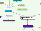
Compartments of the Leg
Lateral Compartment
Superior/Inferior Retinaculum
Fibular Artery Branch
Superficial Fibular Nerve
(L5, S1, S2)
Fibularis Longus
Fibularis Brevis
Anterior Compartment
Retinaculum
Superior/ Inferior External Retinaculum
Innervation:
Anterior Tibial Vein
Anterior Tibial Artery
Deep Fibular Nerve
L4-L5
Mneumonic: FEET
Extensor Hallucis Longus
Extends great toe and dorsiflexes ankle
Extensor Digitorum Longus
Extends lateral four digits and dorsiflexes ankle
Tibialis Anterior
Dorsiflexes ankle and inverts foot
Posterior Compartment
Flexor Retinaculum
Superficial Posterior Compartment
Plantaris
Weakly assists gastroc in PF ankle
Soleus
Plantarflexes ankle independent of position of knee
Steadies leg on foot
Gastrocnemius
Flexes leg at knee joint
Raises heel during walking
Plantarflexes ankle when knee is extended
Deep Posterior Compartment
Muscles:
Flexor Hallucis Longus
Flexes great toe at all joints
weakly PF ankle
Supports medial longitudinal arch of foot
Flexor Digitorum Longus
Flexes lateral four digits
PF ankle
Supports longitdunial arches of foot
Subtopic


