IN ORGANELLES
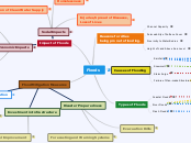
Amino Acids "Tell" proteins where they must go
Called " Membrane Protein"
Further Modification Occurs
Protein folds into 3D shape
Called "Glycosylation"
Endoplasmic Reticulum allows newly made protein to enter the ER lumen
Carbohydrates attach through enzymes
Results in: a glycoprotein
Protein gets packed to a Transport Vesicle
Vesicle delivered to cis side of Golgi
Folded protein gets packed into another transport Vesicle
Chloroplasts
Plastids
Peroxisomes
Can return to ER
Lysosomes
Can travel to Cytoplasm
Folded proteins get packed into secretory Vesicle (specific type) on the Trana side of Golgi
Delivered to Plasma membrane
Embed into Membrane if they are membrane-embedded
Undergo exocytosis if they are free floating
Secrete out of cell
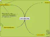
it limits the amount of mRNA that is produced from a particular gene
binding specific transcriptional regulatory proteins to enhancers
in bacteria= same operon system
depending on enviroment it is in
Chromosomes
Activator and enhancer region
7 different control points in eukaryotes
In eukaryote= gametes
Operon system depending on lactose and glucose
regulatory genes (regulators)= affect transcription/ translation in making final protein
All body cells have same chromosomes
regulating by turning off (repressing)
regulating by turning on (inducing)
Organic Molecules
Cellular Respiration Formula
C6H12O6+6O2=6CO2+6H2O+ATP
Goes through 3 stages
Krebs Cycle
Last step in cellular respiration
2 ATP and NADH made
Glycolysis then converted to pyruvate
Glucose
Primary Reactant
Oxidized
Oxygen
Reduced
Used in Cellular Respiration
Occurs in mitochondria
Produces ATP
Energy for the body
Metabolic Pathway
Oxygen is used as the electron acceptor
Used for the ETC
2 Molec. of pyruvate from glycolysis
Subtopic
Happens in Cytosol
USED: 2 ATP
FORMED: 4 ATP, 2 NADH
NET: 2 ATP , 2 Pyruvate molec. , 2 H2O , 2 NADH
Step 4:
Fructose 1,6 bisphosphate--->TWO Glyceraldehyde-3-Phosphates(G3P)
Enzyme in use:Phosphofructokinase/PFK
What it does:Transfers phosphate group FROM ATP to opposite end of sugar (USING 2ND MOLEC. OF ATP)
ATP--->ADP
(Using phosphate group from ATP)
Step 3:
Fructose 6 phosphate--->Fructose 1,6 bisphosphate
Enzyme in use:
Phosphoglucoisomerase
What it does:Converts glucose 6P to fructose 6P
Step 2:
Glucose 6-Phosphate--->Fructose 6 phosphate
ATP ---> ADP
(Using phosphate group from ATP)
Enzyme in use: Hexokinase
What it does:Transfers phosphate group from ATP to glucose
Step 1: Glucose-->Glucose 6 Phosphate
Hydrogen Bonds Link Adenine and Thymine together in DNA. And they line Adenine and Uracil in RNA
Glycolysis
Aerobic Cell Respiration
This unique arrangement acts as a formidable barrier to the free passage if polar molecules and ions.
Pyruvate Oxidation
Polar v Nonpolar Molecules
Nonpolar Molecules (O + CO2): they can easily diffuse the lipid bilayer because they are soluble in the hydrophobic interior.
Polar Molecules (Glucose + Ions): Struggle to pass through the lipid bilayer because of their interaction with the hydrophobic region.
Hydrogen bonds link Uracil and Adenine together in RNA
Hydrogen Bonds link adenine and thymine together in DNA
Hydrogen Bonds join these (G + C) together.
Chemical Bonds
Cell Communication
G Protein Coupled Receptor
cAMP is a second messenger in signaling pathway of a G protein
GCPR is activated with a first messenger bind
Then the now activated GCPR binds to the G protein then activates GTP and activated G protein.
Attaches to Adenyl Cyclase which causes phosphatase to occur
Activated Adenyl Cyclase converts cAMP to ATP
Then second messenger cAMP activates another protein
Cellular Response
Tyrosine Kinase
Inactive monomers with a ligand binding site
When signal molecules bond with receptor sites they create dimer of two tyrosine kinase receptors
Dimerization activates phosphorylation
The active receptor is now recognized by multiple relay proteins
Cellular Response
Energy Transfer
Cell Membranes as Permeable Barriers
Cell membranes are selectively permeable barriers due to their lipid bilayer and the presence of various proteins and cholesterol. They serve as a protective boundary that separates the interior of a cell from its external environment. This selectively permeability is crucial for a variety of cellular functions. As a result of these properties, the membranes can regulate the passage of ions, nutrients, waste products, and other molecules in and out of the cell. This ensures that the internal environment remains stable and conducive for the cell's ideal functioning. This selective permeability is vital for processes like nutrient uptake, waste removal, signal transduction, and the maintenance of cellular homeostasis.
Fluid Mosaic Model
Cholesterol
Interspersed within the lipid bilayer
They prevent the membrane from becoming too rigid. This dynamic adjustment of fluidity ensures that the membrane functions optimally under varying environmental conditions.
Present in the cell membrane, they help regulate the fluidity and stability of the membrane.
Glycoproteins + Glycolipids
Proteins and lipids with attached carbohydrate chains. They play the roles in cell recognition, adhesion, and signaling.
Proteins
These are highly selective and facilitate the transport ions, nutrients, and other needed substances. Contains integral and peripheral proteins. These integral proteins traverse the lipid bilayer, they can have channels or transporters that allow specific molecules to pass through the membrane.
Transport Proteins
These transport proteins that often require energy, in the case of active transport, or they will rely on the concentration gradients, this in the case of passive transport, to help move substances.
Receptor Proteins
Represents the membrane as a dynamic and constantly changing structure, with lipids and proteins able to move within the bilayer, this further influences permeability.
Proteins in the cell membrane act as receptors for specific molecules, this allows the cell to detect and respond to environmental signals.
Lipid Bilayer
This is the basic structure of the cell membrane.
These lipids have hydrophobic tails (water repelling) and hydrophilic heads (water loving). These create a barrier to the passage of water-soluble substances, as they repelled by the hydrophobic core of the membrane.
The hydrophobic tails face towards the interior of the bilayer.
The hydrophilic heads face the aqueous environment both in and outside the cell.
Composed primarily of phospholipids.
Selectively Permeable
Regulating Homeostasis
Osmoregulation: defined as the control of water balance; nutrient uptake, waste elimination, and maintaining an electrochemical gradient across the membrane
Cells must control the passage of ions and molecules to maintain the proper concentration of substances inside and out.
Achieves through combination of factors including the bilayer and various other proteins embedded within the membrane.
Allows positive, and necessary substances to pass through while also preventing unnecessary substances from entering.
Ionic Bonds
Ionic bonds are formed between two or more atoms by the transfer of one or more electrons between atoms. Electron transfer produces negative ions called anions and positive ions called cations.
Hydrogen Bond
Interaction involving a hydrogen atom located between a pair of other atoms having a high affinity for electron; such a bond is weaker than an ionic bond or covalent bond but stronger than van der Waals forces.
covalent bonds
Polar Covalent bond
A bond where electrons are unequally shared by the atoms and spend more time close to one atom than the other.
Because of the unequal distribution of electrons between the atoms of different elements, slightly positive and slightly negative charges develop in different parts of the molecule
Non Polar Covalent Bond
A bond between two atoms of the same element, or between atoms of different elements that share electrons more or less equally.
Cells
Eukaryotic Cells
Centriole
Function: Play the role of organizing microtubules so that they can serve their functions as the cell's skeletal system. They help to determine the locations of the nucleus and other organelles in the cell.
Structure: 9 circularly arranged triplet microtubules. There paired barrel shaped organelles located in the cytoplasm of animal cells near the nuclear envelope.
Smooth ER
Function: Responsible for the synthesis of the essential lipids like phospholipids and cholesterol. Its also responsible for the production and secretion of steroid hormones and the metabolism of carb. Lastly, it stores and releases calcium ions.
Structure: Tube like structure located near the cell periphery. These tubes sometimes branch and form a network that is reticular on first glance. The network of smooth ER allows an increased surface area to only have the purpose of storage to key enzymes
Golgi Apparatus
Function: Works as a factory where proteins coming from the ER are processed and sorted for transport to their designated destinations. As well as synthesizing glycolipids and sphingomyelin within the golgi
Structure: Stack of flattened cisternae and associated vesicles. This transfers proteins and lipids coming from the ER from the cis face and exiting through the trans face.
Mitochondria
Function: The powerhouse of the cell. Generates most of the chemical energy needed to power the biochemical cell reactions.
Structure: Has its own double membrane with inner and outer mitochondrial membranes, and is separated by an intermembrane.
Nucleus
Function: The respiratory of genetic information and is the cell's control center. DNA replication and transcription, as well as RNA processing all take place within the nucleus.
Structure: Lives in the double membraned organelle, withholds the nucleolus, holds 25% of volume. Chromatins are found within the nucleus containing proteins and DNA
Rough ER
Function: Produces/Transfers proteins throughout the rest of the cell to function. Its ribosomes are small and round whose function is to make those proteins
Structure: Flattened membrane sheets (Cisternae), they surround a part of the nucleus and extend across the cytoplasm. Sections of this Cisternae has ribosomes attached and held together by microtubules of the cytoskeleton.
Organelles in Common
Cell Membrane
Function: Manages the transport of materials entering and exiting the cell. They keep toxic substances out of the cell. They contain receptors and channels that allow specific molecules (ions, nutrients, wastes, and metabolic products), mediate cellular and extracellular activities passing between organelles. Lastly, they maintain cell mobility, secretions, and absorptions of substances.
Structure: The main part of the structureis the phospholipid bilayer which forms a stable barrier between 2 compartments. Inside the plasma membrane, these compartments are the in and outside of the cell.
Ribosomes
Function: The ribosome is the sole site for protein synthesis. They read the mRNA sequence and translate the genetic code into an amino acids string which lead to grow into long chains that fold to form proteins. Attaches itself to Rough ER when available (Only in Euk)
Structure: Each ribosome consists of 2 separate RNA-protein complexes (aka small and large subunits). The ribosomes themselves are made up of RNA and proteins.
Cytoplasm
Function: This holds all the works for cell expansion, cell growth, and cell replication throughout it. While doing this, it also protects the cells from damage.
Structure: Gel like fluid inside the cell. It provides a sort of like a stabilization to keep all the other organelles in place, which gives the cell itself its shape.
Prokaryotic Cells
Nucleoid
Function: for controlling the activity and reproduction of the cell. In the nucleoid, it's the place where transcription and replication of DNA takes place. It binds proteins leading them to have a lower molecular mass. This alters the shape of the nucleoid itself.
Structure: Largely composed of about 60% DNA, plus a part of it is made up of RNA and protein. It is proven to had a defined, and self-adherent shape and an underlying longitudinal shape. It lacks membrane and is located within the cytoplasm. It contains proteins, including enzymes and RNA as well as DNA.
Flagella
Structure: A coiled, string-like structure that's sharp bent and consists of a rotary motor at its base and are composed of the protein flagellin. The basal body, a rod and a system of rings embedded in the cell envelope; the hook, which functions as a universal joint; and the filament, composed of thousands of copies of protein flagellin arranged helically and ending with a filament cap composed of an oligomer (a molecule that consists of few repeating units which could be derived, actually or conceptually from molecules)
Function: Gives motility organelle that enables movement and chemotaxis. They spin around and propel the cells very quickly. They act as little fins or tails to help the cell move.
Pili
Function: They initiate contacts between mating pairs. They also facilitate the transfer of genetic material and they draw mating cells into close contact which increased the fertility of the union. They also have a role in the movement process but are most commonly involved with the adherence to surfaces which facilitates infection and is the key to virulence characteristic.
Structure: Resembles hair attached to the outside of the prokaryote (attached to cell wall). It's typically associated with bacterial adhesion related to bacterial colonization and infection. They are primarily composed of oligomeric pili proteins, which arrange helically to form a cylinder. When new pili protein molecules are made, they insert into the base of the pilius.
Plasmid
Function: They're used in this genetic engineering to amplify as many copies of genes as possible. They are typically tools of cloning, transferring, and manipulating genes.
Structure: A small, circular, double stranded DNA molecule that's distinct from a cell's chromosomal DNA. They naturally exist in bacterial cells. These genes often carry the plasmids provide bacteria with genetic advantages, like antibiotic resistance. Most commonly found in the cytoplasm.
Cell Wall
Function: While the cell wall surrounds the plasma membrane of plant cells, it also provides a tensile like strength and protection against mechanical and osmotic stress and other enemies that reveal as a threat to the cell. It also allows the cell to develop turgor pressure which is the pressure of the cell contents against the wall. It's basically a wall meant to protect all the components of the prokaryotic cell.
Structure: The cell wall is one of the outermost layers of the cell. It's composed of cellulose microfibrils and cross linking glycans which are embedded in a highly cross-linked matric of pectin polysaccharides.
Capsule
Function: This helps prokaryotes attach to each other and to various other surfaces in their environment, it also helps prevent the cell from drying out. It's also a sort of boundary which protects bacteria from toxic compounds and desiccation (drying out) and allows them to again adhere to surfaces and escape the immune system of the host.
Structure: It's the sticky, outermost layer of the cell which is usually made of polysaccharides.
DNA & RNA Monomers
Both
Cytosine
FUNCTION: codes genetic information that molecules carry. It can be made into different bases to carry epigenetic information. Energy carrier and cofactor of CTP. Paired with Guanine(4/4)
STRUCTURE: a single heterocyclic aromatic ring, a keto group of C2 and an amino group at C4c. It's molecule is a planar shape. Cytosine can form 3 hydrogen bonds with guanine
Guanine
FUNCTION: A building block of DNA which encodes information. It's also used for energy in G proteins. Paired with Cytosine. (3/4)
STRUCTURE: Composed of 2 nitrogen, 3 carbon, 4 hydrogen, and 1 oxygen. It's 2 rings attached to each other, 5 membered ring and a 6 membered ring.
Adenine
FUNCTION: A nitrogenous bases for the nucleic acid synthesis. 1/2 purine nucleobases, it's job is to produce nucleotides for nucleic acids. It's typically paired with Thymine. Belongs to nucleotide group called purines (1/4)
STRUCTURE: Made of carbon, nitrogen, and hydrogen atoms. It's chemical formula is C5H5N5. When it attaches to ribose and phosphate, it forms a nucleotide.
DNA
Thymine
FUNCTION: Helps stabilize nucleic acid structures. Paired with Adenine. (2/4)
STRUCTURE: Composed of a single ring consisting of carbon (4), hydrogen (5), nitrogen (2), and oxygen (2).
RNA
Uracil
FUNCTION: Uracil (RNA) replaces Thymine (DNA) in RNA. It helps carry out enzyme synthesis that's necessary for cells to function through bonding with the riboses and phosphates
STRUCTURE: 4 hydrogen, 4 carbon, 2 nitrogen, and 2 oxygen. It's atoms that are bonded together, gives a ring shape.
Translation
tRNA
Amino Acyl tRNA Synthetase
Adds amino acids to 3' of tRNA
Ribosomes
Eukaryotes
80's
Binding Sites
EPA
Prokaryotes
70's
Formation of Proteins from mRNA
In cytoplasm
Forms polypeptide chains
consists of amino acids
Peptidyl Transferase
Makes peptide bonds
Stop Codons
UAA, UGA, UAG
Start Codon: AUG
Prokaryotes: F- Met
Eukaryotes: Met
Transcription
Main enzyme: RNA Polymerase
Synthesizes 5' to 3'
Reads 3' to 5'
Initiation Phase
Makes mRNA
Pre mRNA in Eukaryotes
Poly-A Tail on 3'
Protects from degradation in cytosplasm
uses ATP
Introns and Exons
Spliceosomes
Introns Removed
Mature mRNA
mRNA leaves the nucleus
Modified Guanine 5'Cap
Allows Translation to occur
Recognition signal for Ribosomes to bind
Upstream
Promoter
TATA Box
DNA Sequence
Protein Pathway(s)
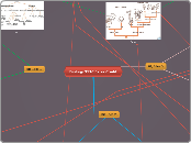
DNA Replication Mechanism
Conservative
2 parental strands reassociate after acting as templates for new strands, this restores the original parental double helix
This visualization is a lot easier because it can only have 2 options: 2 pure dna strands. 3 pure light blue DNA and 1 pure dark blue DNA
Dispersive
each strand both daughter molecules contains a mixture of old and newly synthesized
A way to visualize the dispersed model is the result in the first replication being unpredictive and messy strands. very messy strands, unpredictable
Semi-Conservative
2 strands of the parental molecule separate and each functions as a template for synthesis of a new complementary strand
A way to visualize in 1 pure strand separates and in the first replication you will end up with 2 strands of DNA equally dispersed into it. the 2nd replication could result in 2 equally split DNA strands together and 2 pure strands of DNA. 1 light blue and 1 dark blue strand combined and a pure light blue strand of DNA
