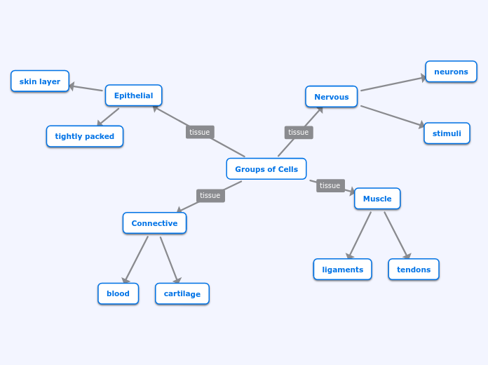
There are various types of tissue in the body, each with distinct functions, locations, and appearances. Nervous tissue, found in the spinal cord and brain, is responsible for receiving stimuli and transmitting impulses.

Location: In peripheral nerves and in organs of the central nervous system, brain, and spinal cord.
Function: Receives stimuli and sends impulses to the spinal cord and Brain.
Appearance: Consists of neurons, dendrites, and axons that form a long line of connectivity.
Muscle
Skeletal Muscle
Location: Attached to the Skeleton.
Function: Moves the body.
Appearance: Is striated and conatins long, thin, multi nucleated fibres that are crossed with a regular pattern of fine red and white lines.
Cardiac Muscle
Location: Walls of the heart.
Function: Controls the contraction and relaxation of the heart.
Appearance: Contains bundles that are branched like a tree but is connected at both ends.
Smooth Muscle
Location: In the walls of hollow visceral organs.
Function: Controls involuntary movements.
Appearance: Contains small smooth muscle cells that our spindle shaped and have no striations but have bundles of thin and thick filaments.
Connective
Bone Tissue
Location: Throughout the body.
Function: Locomotion, support and protection of soft tissues, calcium and phosphate storage, and harboring of bone marrow.
Appearance: Concentric rings of matrix that surround central canals that contain blood vessels that inside contain lacunae that are connected to each other through canalicula.
Blood Tissue
Location: Around every blood vessel.
Function: Transports Oxygen and Carbon Dioxide.
Appearance: Contains a fluid matrix and no fibers.
Fibrocartilage
Location: Between Vertebrae
Function: Shock absorber
Appearance: Tough, dense, and fibrous material.
Elastic Cartilage
Location: External Ear and Larynx.
Function: Greatly flexible to efficiently channel sound waves to the inner ear.
Appearance: Contains many yellow elastic fibers with chondrocytes that lie between them that appear dark.
Hyaline Cartilage
Location: Ends of joints, nose and respiratory passages.
Function: Shock absorber and reduces friction by serving as a padding between bones where they meet at joints.
Appearance: Glass-like but translucent with a pearl-grey color and a considerable amount of collagen with a firm consistency.
Fibrous
Location: Dermis of skin, tendons, and ligaments.
Function: Support
Appearance: Large amounts of collagen fibers and few cells or matrix material.
Adipose
Location: Subcutaneous fat, visceral fat, bone marrow, and breasts.
Function: Serves as a protective cushion, insulation to preserve body heat, and stores energy.
Appearance: Loose connective tissue composed of adipocytes.
Loose Connective or Areolar Tissue
Location: Skin, around mucous membranes, around blood vessels, nerves, and organs of the body.
Function: Binds underlying organs to skin and to each other and also forms delicate thin membranes throughout the body.
Appearance: Loose arrangement of collagenous and elastic fibers, scattered cells of various types; abundant ground substance; numerous blood vessels
Epithelial
Stratified Columnar
Location: Parts of the Eye, Pharynx, Anus, Uterus, Male Urethra, and Vas Deferens