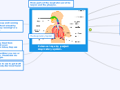
CARDIOPULMONARY
ANATOMY
CARDIOVASCULAR
SYSTEM
To help maintain fluid balance within the body
To help the body maintain a constant body temperature
To protect
the body from infection and blood loss
To transport
nutrients, gases and waste products around the body
LYMPHATIC
SYSTEM
LYMPH
NODES
filter lymph fluid before returning it to the blood
SPLEEN
it controls the amount of red blood cells and blood storage in the body
THYMUS
hematopoietic precursors from the bone-marrow
LYMPH
transports oxygen, food materials, hormones
transports white blood cells to and from the lymph nodes
It absorbs and transports fatty acids
removal of interstitial fluid from tissues.
BLOOD
VESSELS
VEINS
VENULES
serve to return blood from organs to the heart
ARTERY
COMPONENTS
CAPILLARIES
ARTERIOLES
vessels that carry blood oxygenateda way from the heart
Water balance
PH
Transport
Hormones
Nutrients
Gases (O2 and CO2)
BLOOD
FORMED ELEMENTS
PLATELETS
are pushed out from the center of flowing blood to the wall of the blood vessel
LEUKOCYTES
are actively engaged in the destruction or neutralization of invading microorganisms
ERYTHROCYTES
transportation of oxygen and carbon dioxide
PLASMA
transport nutrients throughout the body
Protection
Regulation
Transportation
HEART
FOUR CHAMBERS
LEFT VENTRICLE
Pumps oxygenated blood from lungs to body
RIGHT VENTRICLE
is the chamber within the heart that is responsible for pumping oxygen-depleted blood to the lungs.
LEFT ATRIUM
It plays the vital role of receiving blood from the lungs via the pulmonary veins and pumping it to the left ventricle.
RIGHT ATRIUM
which function as receiving chambers for blood entering the heart
PERICARDIUM
Lubricates the heart
Prevents excessive dilation of the heart in cases of acute volume overload
Protects it from infections coming from other organs
Sets heart in mediastinum and limits its motion
It takes in deoxygenated blood through the veins and delivers it to the lungs for oxygenation before pumping it into the various arteries
Week 8
Day 50
aorta - pulmonary trunk are already separated
Week 7
Day 49
absorption pulmonary veins
Four-chambered heart
Week 6
Day 42
Division of truncus arteriosus
Day 40
formation bulb-ventricular septum
Day 39
full bottom wall
Day 36
Septum secundum
Week 5
Day 35
three-chambered heart
Atrioventricular orifice
Day 31
septum primum
Day 30
circulation starts
Day 29
Lobed atrium
Week 4
Day 26
single atrium
Day 25
cardiogenic loop is formed
Day 23
first contraction
Only half a heart tube
Day 22
Fusion endocardial tubes
Week 3
Day 21
Endocardial tubes appear
Day 20
There cardiogenic plate
PULMONARY
SYSTEM
Olfaction, or Smelling, Is a Chemical Sensation
Air Vibrating the Vocal Cords Creates Sound
Internal Respiration Exchanges Gases Between the Bloodstream and Body Tissues
External Respiration Exchanges Gases Between the Lungs and the Bloodstream
Inhalation and Exhalation Are Pulmonary Ventilation—That’s Breathing
STRUCTURES
DIAPHRAGM
is the main respiratory muscle that allows us to inhale and exhale
ALVEOLI
Gas exchange of oxygen and carbon dioxide takes
MAIN BRONCHI
When someone takes a breath through their nose or mouth, the air travels into the larynx
LOBAR
BRONCHUS
LEFT INFERIOR
LEFT SUPERIOR
RIGHT INFERIOR
RIGHT MIDDLE
RIGHT SUPERIOR
LUNGS
distributed to tissues all over the body and expel carbon dioxid
LEFT
TWO LOBES
RIGHT
THREE LOBES
INFERIOR
MIDDLE
SUPERIOR
CARINA'S
TRACHEA
the site of the tracheal bifurcation at the lower end of the trachea
TRACHEA
uns down behind the breastbone
begins just under the larynx
EPIGLOTTIS
also protects the body from choking on food that would normally obstruct the airway.
PHARYNX
It directs the passage of air and food between the mouth and nose into esophagus and larynx
NASAL CAVITY
FUNCTION
condition the air to be received by the other areas of the respiratory tract
EMBRYOLOGY
Hypertrophy 3-8 years
Increased cell size
Active hyperplasia 0-3 years
Increases the number of alveoli
Microvascular maturation 0-3 years
fusion of a unique capillary bilayer
Interdental wall thinning
Alveolar 36 weeks and finished
alveoli formation
Development of secondary septa
Saccular 28-35 weeks
Training saccules
Formation of the transitional airspace
Canalicular 17-27 weeks
It appears surfactant
epithelial differentiation
Growth of the capillary bed
Forming acini
Pseudoglandular 7-17 weeks
vascular growth
Development of the bronchial tree to level
terminal bronchioles
Occurrence of pulmonary circulation
Embryonic 3-6 weeks
Day 37
The
lobar bronchi begin their training
Day 33
the division occurs in two branches
Main and lung buds
development of large airways
It originates from the mesoderm
